
Page 19 - Libro 2
P. 19
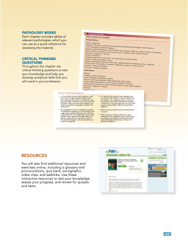
    PATHOLOGY BOXES
Each chapter includes tables of relevant pathologies, which you can use as a quick reference for reviewing the material.
CRITICAL THINKING QUESTIONS
Throughout the chapter are critical thinking questions to test your knowledge and help you develop analytical skills that you will need in your profession.
                          Critical Thinking Questions
1. A 3-year-old boy presents with hematuria, a mild fever, and a left upper quadrant palpable mass.
The sonogram demonstrates a well-defined, homo- geneously solid, 3-cm mass in the lower pole of the left kidney. What is the most likely diagnosis and where else should the sonographer include in the examination?
2. The sonographer receives a requisition to perform an abdominal sonogram on a 2-day-old infant with a right upper quadrant abdominal mass following a difficult delivery. The examination reveals a large echogenic mass superior to the right kidney and appears separate from the right kidney. What is the most likely diagnosis and what would help to confirm the diagnosis?
3. A 6-month old girl presents with a palpable mass just inferior to the umbilicus. The area appears red and inflamed. The sonogram reveals a cystic mass that contains some debris. With further evaluation, a small tract is seen connecting the cystic area to the superior urinary bladder. What is the most likely diagnosis?
4. What is the most common cause of hydronephrosis in infants?
5. While scanning a newborn patient for a renal examination, the sonographer notices both kidneys are enlarged and echogenic with hyperechoic foci scattered throughout both kidneys. What is the most likely diagnosis?
           RESOURCES
You will also find additional resources and exercises online, including a glossary with pronunciations, quiz bank, sonographic video clips, and weblinks. Use these interactive resources to test your knowledge, assess your progress, and review for quizzes and tests.
  xvii
 A
o
n
c
c
c
i
t A
p
r
n
r
u
t
n
s
r
p
r
c
PATHOLOGY BOX 20-1
Causes of Renal Size Variation
Enlarged Kidneys
Bilateral enlargement
Congenital: Duplication, cystic disease, storage disease, generalized visceromegaly, systemic infection
Acute: Pyelonephritis, glomerular nephritis
Neoplastic: Nephroblastomatosis, bilateral Wilms tumor, leukemia, lymphoma, tuberous sclerosis, or hamartoma Vascular: Renal vein thrombosis, acute tubular necrosis, hemolytic uremia, sickle cell anemia
Obstructive: Congenital or acquired
Unilateral enlargement
Congenital: Duplication, cystic disease, cross-fused ectopia, horseshoe kidney
Infectious: Acute pyelonephritis, abscess
Neoplastic: Mesoblastic nephroma, Wilms tumor, angiomyolipoma or hamartoma, sarcoma, lymphoma Vascular: Renal vein thrombosis, transplant complication (rejection or tubular necrosis)
Traumatic: Contusion, hematoma
Obstructive: Congenital, acquired
Small Kidneys
Bilateral
Congenital: Aplasia, hypoplasia
Acute: Pyelonephritis, glomerular nephritis
Infectious: Chronic pyelonephritis, reflux nephropathy with infarction
Vascular: Renal vein thrombosis, arterial occlusion (intrinsic or extrinsic)
Atrophic: Chronic obstruction, chronic recurrent infarction, chronic failure, dysplasia Obstructive: Congenital or acquired
Unilateral
Congenital: Agenesis, hypoplasia
Infectious: Chronic, chronic reflux with infection
Vascular: Venous thrombosis, arterial obstruction (acquired or congenital)
o
n
a
h
h
i
i
c
:
C
:
C h
o
c i
o b
o
r
n
h
r
o
i
c
o
b
s
r
t
s
t
u
a
t
c
,
h
c
n
n
t
,
c h
o
i
i
o
survive for only a very short time. They may live longer with v til
h d
r
o
n
i
i
i
o n
c
i
n
f
e
t
e f
c t
i
s i
i
o
n
a
n
a
t
d
i
d
i
n
n
f
f
r
t
a
c
i
a
r
c
i
o
o
n
n
,
d
,
d
20 — The Pediatric Urinary System and Adrenal Glands 5
y
y
s
p
s
p
l
a
l
a
