
Page 185 - Libro 2
P. 185
10 — Upper Extremity Arterial Duplex Scanning 165
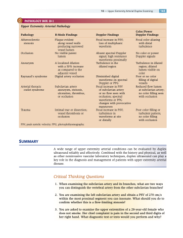
PATHOLOGY BOX 10-1
Upper Extremity Arterial Pathology
Color/Power Pathology B-Mode Findings Doppler Findings Doppler Findings
Atherosclerotic stenosis
Occlusion Aneurysm
Raynaud’s syndrome
Arterial thoracic outlet syndrome
Trauma
Plaque evident
along vessel walls producing narrowed vessel lumen
No visible patent lumen
A localized dilation with a 50% increase as compared to the adjacent vessel
Digital artery occlusion
Subclavian artery aneurysm, stenosis, ulceration, thrombus, or occlusion
Intimal tear or dissection; vessel thrombosis or occlusion
Focal increase in PSV; loss of multiphasic waveform
Absent spectral Doppler signal; high resistance waveforms proximally
Turbulence in the dilated region
Diminished digital waveforms on spectral Doppler or PPG
Focal increase in PSV
of subclavian artery
or no flow seen with occlusion; spectral waveforms or PPG changes with provocative maneuvers
Focal increase in PSV; turbulence in waveforms at site or distally
Focal color aliasing with distal turbulence
No color or power Doppler signals
Turbulence in dilated region; dilated lumen visible on color
Poor or no color filling of digital vessels
Reduced flow lumen at subclavian artery; no color filling seen with occlusion
Poor color filling or turbulent pattern; no color filling with occlusion
PSV, peak systolic velocity; PPG, photoplethysmography.
SUMMARY
A wide range of upper extremity arterial conditions can be evaluated by duplex ultrasound reliably and effectively. Combined with the history and physical, as well as other noninvasive vascular laboratory techniques, duplex ultrasound can play a key role in the diagnosis and management of patients with upper extremity arterial disease.
Critical Thinking Questions
1. When examining the subclavian artery and its branches, what are two ways you can distinguish the vertebral artery from the other subclavian branches?
2. You are examining the left subclavian artery and obtain a PSV of 270 cm/s within the most proximal segment you can insonate. What should you do to confirm whether this is a flow-limiting stenosis?
3. You are asked to examine the upper extremities of a 28-year-old female who does not smoke. Her chief complaint is pain in the second and third digits of her right hand. What diagnostic test or tests would you perform and why?
