
Page 184 - Libro 2
P. 184
164
PART 3 — PERIPHERAL ARTERIAL
as an effective means of evaluating for occult arterial injury. If immediate operative repair is not indicated, positive findings can be followed up with arteriogra- phy. A well-performed normal arterial duplex exami- nation essentially rules out major clinically significant injuries of upper extremity arteries distal to the axil- lary crease. The ultrasound techniques for examining patients with suspected arterial trauma are those used for standard upper extremity evaluation, but additional considerations should be taken. Depending on the type of trauma (particularly a gunshot wound), the arterial injury may be remote to a wound. All vessels in the re- gion should be examined for intimal tears or dissections as these are often associated with traumatic injuries.
ARTERIAL OCCLUSIVE DISEASE
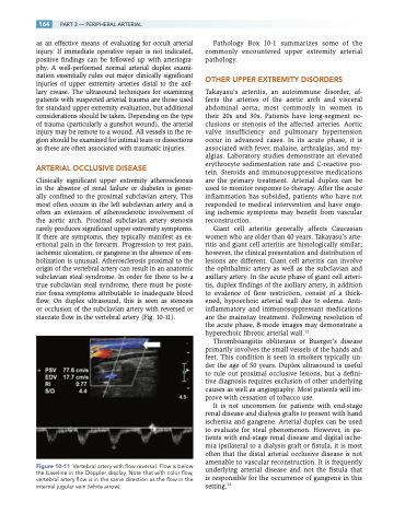
Clinically significant upper extremity atherosclerosis in the absence of renal failure or diabetes is gener- ally confined to the proximal subclavian artery. This most often occurs in the left subclavian artery and is often an extension of atherosclerotic involvement of the aortic arch. Proximal subclavian artery stenosis rarely produces significant upper extremity symptoms. If there are symptoms, they typically manifest as ex- ertional pain in the forearm. Progression to rest pain, ischemic ulceration, or gangrene in the absence of em- bolization is unusual. Atherosclerosis proximal to the origin of the vertebral artery can result in an anatomic subclavian steal syndrome. In order for there to be a true subclavian steal syndrome, there must be poste- rior fossa symptoms attributable to inadequate blood flow. On duplex ultrasound, this is seen as stenosis or occlusion of the subclavian artery with reversed or staccato flow in the vertebral artery (Fig. 10-11).
Figure 10-11 Vertebral artery with flow reversal. Flow is below the baseline in the Doppler display. Note that with color flow, vertebral artery flow is in the same direction as the flow in the internal jugular vein (white arrow).
Pathology Box 10-1 summarizes some of the commonly encountered upper extremity arterial pathology.
OTHER UPPER EXTREMITY DISORDERS
Takayasu’s arteritis, an autoimmune disorder, af- fects the arteries of the aortic arch and visceral abdominal aorta, most commonly in women in their 20s and 30s. Patients have long-segment oc- clusions or stenosis of the affected arteries. Aortic valve insufficiency and pulmonary hypertension occur in advanced cases. In its acute phase, it is associated with fever, malaise, arthralgias, and my- algias. Laboratory studies demonstrate an elevated erythrocyte sedimentation rate and C-reactive pro- tein. Steroids and immunosuppressive medications are the primary treatment. Arterial duplex can be used to monitor response to therapy. After the acute inflammation has subsided, patients who have not responded to medical intervention and have ongo- ing ischemic symptoms may benefit from vascular reconstruction.
Giant cell arteritis generally affects Caucasian women who are older than 40 years. Takayasu’s arte- ritis and giant cell arteritis are histologically similar; however, the clinical presentation and distribution of lesions are different. Giant cell arteritis can involve the ophthalmic artery as well as the subclavian and axillary artery. In the acute phase of giant cell arteri- tis, duplex findings of the axillary artery, in addition to evidence of flow restriction, consist of a thick- ened, hypoechoic arterial wall due to edema. Anti- inflammatory and immunosuppressant medications are the mainstay treatment. Following resolution of the acute phase, B-mode images may demonstrate a hyperechoic fibrotic arterial wall.12
Thromboangiitis obliterans or Buerger’s disease primarily involves the small vessels of the hands and feet. This condition is seen in smokers typically un- der the age of 50 years. Duplex ultrasound is useful to rule out proximal occlusive lesions, but a defini- tive diagnosis requires exclusion of other underlying causes as well as angiography. Most patients will im- prove with cessation of tobacco use.
It is not uncommon for patients with end-stage renal disease and dialysis grafts to present with hand ischemia and gangrene. Arterial duplex can be used to evaluate for steal phenomenon. However, in pa- tients with end-stage renal disease and digital ische- mia ipsilateral to a dialysis graft or fistula, it is most often that the distal arterial occlusive disease is not amenable to vascular reconstruction. It is frequently underlying arterial disease and not the fistula that is responsible for the occurrence of gangrene in this setting.13
