
Page 182 - Libro 2
P. 182
162
PART 3 — PERIPHERAL ARTERIAL
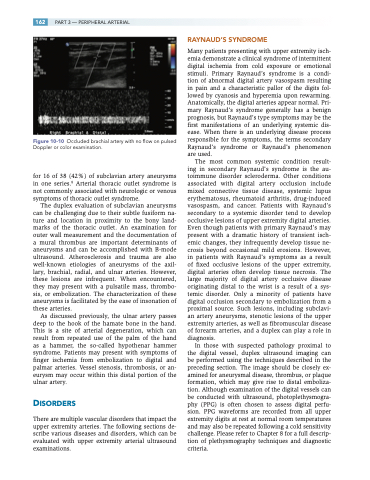
Figure 10-10 Occluded brachial artery with no flow on pulsed Doppler or color examination.
for 16 of 38 (42%) of subclavian artery aneurysms in one series.8 Arterial thoracic outlet syndrome is not commonly associated with neurologic or venous symptoms of thoracic outlet syndrome.
The duplex evaluation of subclavian aneurysms can be challenging due to their subtle fusiform na- ture and location in proximity to the bony land- marks of the thoracic outlet. An examination for outer wall measurement and the documentation of a mural thrombus are important determinants of aneurysms and can be accomplished with B-mode ultrasound. Atherosclerosis and trauma are also well-known etiologies of aneurysms of the axil- lary, brachial, radial, and ulnar arteries. However, these lesions are infrequent. When encountered, they may present with a pulsatile mass, thrombo- sis, or embolization. The characterization of these aneurysms is facilitated by the ease of insonation of these arteries.
As discussed previously, the ulnar artery passes deep to the hook of the hamate bone in the hand. This is a site of arterial degeneration, which can result from repeated use of the palm of the hand as a hammer, the so-called hypothenar hammer syndrome. Patients may present with symptoms of finger ischemia from embolization to digital and palmar arteries. Vessel stenosis, thrombosis, or an- eurysm may occur within this distal portion of the ulnar artery.
DISORDERS
There are multiple vascular disorders that impact the upper extremity arteries. The following sections de- scribe various diseases and disorders, which can be evaluated with upper extremity arterial ultrasound examinations.
RAYNAUD’S SYNDROME
Many patients presenting with upper extremity isch- emia demonstrate a clinical syndrome of intermittent digital ischemia from cold exposure or emotional stimuli. Primary Raynaud’s syndrome is a condi- tion of abnormal digital artery vasospasm resulting in pain and a characteristic pallor of the digits fol- lowed by cyanosis and hyperemia upon rewarming. Anatomically, the digital arteries appear normal. Pri- mary Raynaud’s syndrome generally has a benign prognosis, but Raynaud’s type symptoms may be the first manifestations of an underlying systemic dis- ease. When there is an underlying disease process responsible for the symptoms, the terms secondary Raynaud’s syndrome or Raynaud’s phenomenon are used.
The most common systemic condition result- ing in secondary Raynaud’s syndrome is the au- toimmune disorder scleroderma. Other conditions associated with digital artery occlusion include mixed connective tissue disease, systemic lupus erythematosus, rheumatoid arthritis, drug-induced vasospasm, and cancer. Patients with Raynaud’s secondary to a systemic disorder tend to develop occlusive lesions of upper extremity digital arteries. Even though patients with primary Raynaud’s may present with a dramatic history of transient isch- emic changes, they infrequently develop tissue ne- crosis beyond occasional mild erosions. However, in patients with Raynaud’s symptoms as a result of fixed occlusive lesions of the upper extremity, digital arteries often develop tissue necrosis. The large majority of digital artery occlusive disease originating distal to the wrist is a result of a sys- temic disorder. Only a minority of patients have digital occlusion secondary to embolization from a proximal source. Such lesions, including subclavi- an artery aneurysms, stenotic lesions of the upper extremity arteries, as well as fibromuscular disease of forearm arteries, and a duplex can play a role in diagnosis.
In those with suspected pathology proximal to the digital vessel, duplex ultrasound imaging can be performed using the techniques described in the preceding section. The image should be closely ex- amined for aneurysmal disease, thrombus, or plaque formation, which may give rise to distal emboliza- tion. Although examination of the digital vessels can be conducted with ultrasound, photoplethysmogra- phy (PPG) is often chosen to assess digital perfu- sion. PPG waveforms are recorded from all upper extremity digits at rest at normal room temperatures and may also be repeated following a cold sensitivity challenge. Please refer to Chapter 8 for a full descrip- tion of plethysmography techniques and diagnostic criteria.
