
Page 180 - Libro 2
P. 180
160
PART 3 — PERIPHERAL ARTERIAL
A
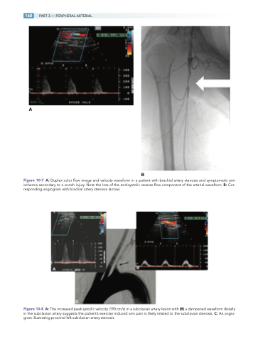
Figure 10-7 A: Duplex color flow image and velocity waveform in a patient with brachial artery stenosis and symptomatic arm ischemia secondary to a crutch injury. Note the loss of the end-systolic reverse flow component of the arterial waveform. B: Cor- responding angiogram with brachial artery stenosis (arrow).
Figure 10-8 A: The increased peak systolic velocity (190 cm/s) in a subclavian artery lesion with (B) a dampened waveform distally in the subclavian artery suggests the patient’s exercise induced arm pain is likely related to the subclavian stenosis. C: An angio- gram illustrating proximal left subclavian artery stenosis
B
