
Page 181 - Libro 2
P. 181
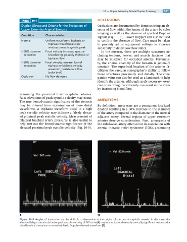
TABLE 10-1
Duplex Ultrasound Criteria for the Evaluation of Upper Extremity Arterial Stenosis
10 — Upper Extremity Arterial Duplex Scanning 161 OCCLUSIONS
Occlusions are documented by demonstrating an ab- sence of flow within the lumen of the artery by color imaging as well as the absence of spectral Doppler signals (Fig. 10-10). Power Doppler can also be used to confirm the absence of flow. Care must be taken to properly adjust equipment settings to increase sensitivity to detect low flow states.
In the forearm, there are multiple structures in- cluding tendons, nerves, and muscle fascicles that may be mistaken for occluded arteries. Fortunate- ly, the arterial anatomy of the forearm is generally constant. The superficial location of the arteries fa- cilitates the vascular sonographer’s ability to follow these structures proximally and distally. The com- panion veins can also be used as a landmark to help identify the arteries. Although rarely necessary, exer- cise or warming the extremity can assist in the exam by increasing blood flow.
ANEURYSMS
By definition, aneurysms are a permanent localized dilation resulting in a 50% increase in the diameter of the artery compared to the diameter of the normal adjacent artery. Several regions of upper extremity arteries deserve consideration. First, aneurysms of the subclavian artery often occur in association with arterial thoracic outlet syndrome (TOS), accounting
Condition
Normal
50% diameter reduction
50% diameter reduction
Occlusion
Characteristics
Uniform waveforms; biphasic or triphasic waveforms; clear window beneath systolic peak
Focal velocity increase; spectral broadening; possibly triphasic or biphasic flow
Focal velocity increase; loss of triphasic or biphasic velocity waveform; poststenotic flow (color bruit)
Noflowdetected
examining the proximal brachiocephalic arteries. False elevations of peak systolic velocity may occur. The true hemodynamic significance of the stenosis may be inferred from examination of more distal waveforms. A triphasic waveform distal to a high peak systolic velocity may indicate a falsely elevat- ed proximal peak systolic velocity. Measurement of bilateral brachial artery pressures is also useful to help sort out the hemodynamic significance of the elevated proximal peak systolic velocity (Fig. 10-9).
AB
Figure 10-9 Angles of insonation can be difficult to determine at the origins of the brachiocephalic vessels. In this case, the elevated left proximal subclavian peak systolic velocity of 247 cm/s (A) may not indicate a hemodynamically significant lesion as the distal brachial artery has a normal triphasic Doppler-derived waveform (B).
