
Page 204 - Libro 2
P. 204
184 PART 3 — PERIPHERAL ARTERIAL
TABLE 12-1
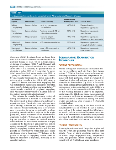
Endovascular Interventions for Lower Extremity Atherosclerotic Occlusive Disease
Intervention Stenosis-Free
Option Mechanism Lesion Anatomy Patency at 1 Year Failure Mode
Balloon angioplasty
Stent angioplasty
Atherectomy
Subintimal angioplasty
Stent-graft angioplasty
Lumen dilation Lumen dilation Plaque excision Lumen dilation Lumen dilation
Focal, 5 cm stenosis or occlusion
Focal and longer (10 cm) stenosis or occlusion
Focal and longer (10 cm) stenosis
10 stenosis or occlusion Long, 15 cm stenosis or
occlusion
40%–50% Plaque dissection Myointimal hyperplasia
50%–60% Myointimal hyperplasia Stent fracture
40%–50% Myointimal hyperplasia Atherectomy site thrombosis
55%–60% Myointimal hyperplasia Angioplasty site thrombosis
60%–70% Myointimal hyperplasia Graft thrombosis
Consensus (TASC II) criteria based on lesion loca- tion and anatomy.4 Endovascular intervention is the preferred therapy for focal, 5 cm in length regions of stenosis or occlusion (TASC A and B lesions) with expected 30-day technical and clinical success rates above 95%.1,4 In claudicants, the patency of iliac an- gioplasty is higher (85% at 3 years) than for super- ficial femoral/popliteal artery angioplasty (55% at 2 years).11,12 Treatment of CLI or TASC C and D lesions by endovascular techniques has lower stenosis-free patency rates, typically in the 25% to 40% range at 1 year.2,8,10,13 Factors associated with angioplasty fail- ure include lesion calcification, occlusions, poor tibial artery runoff, diabetes mellitus, and renal failure.4–10 Approximately one-third of peripheral angioplasty sites will require reintervention to maintain stenosis- free patency during within the first year.7
The rationale for vascular laboratory testing fol- lowing intervention is twofold: to document that the improvement in limb perfusion was sufficient to expect symptoms (claudication, rest pain) and signs (ulcer healing) to resolve and to detect angioplasty- site stenosis. Because the PAD patient is prone to dis- ease progression, including myointimal hyperplasia producing stenosis within or adjacent to the angio- plasty site, duplex ultrasound is the recommended diagnostic modality. Testing can be performed dur- ing the procedure to assess for residual stenosis, which is suited for surveillance studies and is highly accurate in the detection of angioplasty site compli- cations versus ASO progression. Duplex testing can classify angioplasty stenosis severity and thus can provide an opportunity to retreat high-grade occlu- sive lesions prior to thrombosis.7,14,15 Reliance on the patient to recognize angioplasty failure based on symptom recurrence is imperfect, especially in the sedentary patient.
SONOGRAPHIC EXAMINATION TECHNIQUES
PATIENT PREPARATION
Arterial testing after endovascular intervention mir- rors the surveillance used after lower limb bypass grafting.15–18 Patient functional status is documented, including any new or unresolved symptoms of PAD, along with limb perfusion, which is evaluated by physiologic testing and a duplex scan of the angio- plasty site for abnormalities (Fig. 12-1). In the claudi- cant, initial indirect physiologic testing should verify improvement in the ankle–brachial index (ABI) to a normal (0.9) or an increased (0.2) level sufficient so that walking distance is likely to be improved. Limbs treated for CLI should have a measurement of toe pressure to verify that an increase to 30 mm Hg has been achieved. In limbs with healing foot ulcers or digit amputations, a toe pressure of 40 mm Hg predicts healing.
Duplex arterial mapping of the limb should be individualized based on whether the ABI is normal, abnormal, or if the patient has calcified tibial arter- ies, which preclude accurate ankle pressure measure- ment. If the ABI is normal and the tibial artery velocity spectra at the ankle indicate multiphasic or triphasic artery flow, angioplasty site imaging is not necessary.
PATIENT POSITIONING
Scanning is performed with the patient supine and with the lower limb positioned with the knee bent slightly. Prone or lateral decubitus positions can be used to image the popliteal artery, tibioperoneal trunk, and peroneal arteries. Imaging can be per- formed with a 5-7 MHz linear array transducer.
