
Page 206 - Libro 2
P. 206
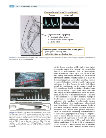
186
PART 3 — PERIPHERAL ARTERIAL
Common Femoral Artery Velocity Sp
Normal Abnormal
ectra
DuplexScan of Angioplasty
A – proximal inflow artery
B – endovascular treated segment C – distal artery
Duplex-acquired ankle-level tibial artery spectra
- peak systolic velocity, PSV
- pulsatility index, acceleration time
Figure 12-2 Schematic-depicting sites of duplex scanning including the common femoral artery, angioplasty site, and assessment of distal tibial artery hemodynamics.
Figure 12-3 Duplex image and velocity spectra of an exter- nal iliac stent stenosis. The color duplex image (top) depicts an area of color aliasing at the stenosis (arrow). The pulsed Doppler (middle) sample volume is walked through the stenosis with a measurement of peak systolic velocity proxi- mal to (PSVprox) and at the site of stenosis (PSVmax) for cal- culation of velocity ratio, Vr PSVmax/PSVprox. A schematic drawing illustrates the stenosis and PSV measurements (bottom).
arterial duplex scanning enable study interpretation in categories ranging from “normal” (i.e., stenosis-free patency) to “severe stenosis,” with the latter category based on threshold criteria appropriate for reinterven- tion. Testing immediately following an endovascular intervention confirms procedural success or failure by documenting patency and whether a residual stenosis is present. Subsequent testing is based on procedure indication; it is less frequent in active individuals treated for claudication, but in patients treated for CLI, surveillance should be similar following lower limb bypass grafting.6 Duplex surveillance after lower limb angioplasty has demonstrated 50% DR steno- sis in 20% to 40% of treated limbs within 12 months of the procedure.7,16 The most common etiology is development of myointimal hyperplasia within the angioplasty site regardless of endovascular therapy (balloon inflation, stent angioplasty, atherectomy) used. Detection of 50% residual stenosis despite a completion angiogram showing adequate lumen ex- pansion (30% DR) can occur, predicts angioplasty, and is the rationale for performing an intraprocedural or early (30-day) postprocedural study.7,16 The preva- lence of duplex-detected residual stenosis following femoropopliteal angioplasty is lowest after stent-an- gioplasty or stent-grafting (5%), and higher after balloon angioplasty (15% to 20%) or atherectomy (25%). Although angiogram-monitored angioplasty showing 20% to 30% residual stenosis predicts
