
Page 207 - Libro 2
P. 207
30-day patency (i.e., technical success), a duplex scan of angioplasty-site hemodynamics provides a more precise assessment of functional patency. Schillinger et al.7 documented that duplex-detected 50% ste- nosis developed more frequently after balloon angio- plasty than after Nitinol stenting in the treatment of SFA occlusive disease at both 6 months (45% vs. 25%, P .06) and 12 months (63% vs. 37%, P .01). Other techniques, including cutting or cryoplasty bal- loon angioplasty, atherectomy, and stent-grafting, also have a high technical success rate, but similar to bal- loon angioplasty, angioplasty restenosis or treatment site thrombosis has been observed in 20% to 50% of limbs depending on TASC-lesion severity by 1 year.11,15
TABLE 12-2
DIAGNOSIS
Peripheral arterial laboratory testing following PTA includes interpretation of the limb pressures and du- plex ultrasound findings (see Fig. 12-1). The inter- pretation should comment on the severity of limb ischemia (mild, moderate, severe), changes from preintervention values, and whether adequate foot perfusion has been achieved in patients treated from CLI (i.e., toe pressure 30 mm Hg). Duplex findings at the angioplasty site are interpreted as showing no stenosis (50% DR stenosis), moderate stenosis (50% DR), severe stenosis (70% DR), or occlu- sion (Table 12-2). A “normal study” interpretation
12 — Ultrasound Following Interventional Procedures 187
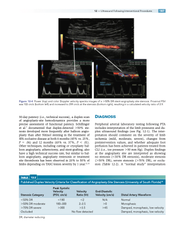
Figure 12-4 Power (top) and color Doppler velocity spectra images of a 50% DR stent-angioplasty site stenosis. Proximal PSV was 103 cm/s (bottom left) and increased to 299 cm/s at the stenosis (bottom right), resulting in a calculated velocity ratio of 2.9.
Published Duplex Velocity Criteria for Classification of Angioplasty-Site Stenosis (University of South Florida)18
50% DR 180 2 N/A Normal
50% DR moderate 180–300 2–3.5 0 Monophasic
70% DR severe 300 3.5 45 Damped, monophasic, low velocity Occluded No flow detected Damped, monophasic, low velocity
Peak Systolic
Velocity Velocity End-Diastolic
Stenosis Category (PSV, cm/s) Ratio (Vr) Velocity (cm/s) Distal Artery Waveform
DR, diameter reducing.
