
Page 209 - Libro 2
P. 209
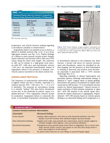
TABLE 12-3
Published Duplex Velocity Criteria for Superficial Femoral Artery Stenosis (University of Pittsburgh)14
Peak Systolic
Stenosis Velocity Velocity
12 — Ultrasound Following Interventional Procedures 189 Sagittal Scan
Transverse Scan
Figure 12-6 Power Doppler images (sagittal, transverse) and an angiogram of a superficial femoral artery stent with diffuse in-stent stenosis. PSV ranged from 200 to 300 cm/s within the stent. Measured ABI was 0.56.
or immediately adjacent to the treatment site. Stent fracture, a known risk factor for stenosis develop- ment and thrombosis, cannot be identified by du- plex imaging, but the presence of stent deformation or kinking is abnormal and predicts stent thrombo- sis especially if associated with a 70% stenosis (Pathology Box 12-1).
Reporting standards of clinical improvement and procedure patency following endovascular therapy is identical to “open” surgical repair or bypass grafting. Intervention site patency documented by duplex ultra- sound and an ABI increase 0.15 are minimal outcome criteria for clinical improvement.3 Clinical success re- quires resolution of limb ischemia symptoms or signs and 50% DR stenosis of the arterial repair docu- mented by duplex ultrasound or angiography. Duplex- detected 50% DR stenosis based on velocity spectra findings is an abnormal finding and if progressive to a
Category
50% DR 50% DR 80% DR Occluded
(PSV, cm/s)
190 190–275 275
Ratio (Vr)
1.5 1.5–3.5 3.5
DR, diameter reducing.
Noflowdetected
progression, and clinical decision making regarding a surveillance schedule or reintervention.
Angioplasty failure can manifest as an occlusion, a diffuse or multiple stenosis (Fig. 12-6), or as a focal, high-grade stenosis (see Fig. 12-5). Duplex findings of a diffuse in-stent stenosis include power Doppler lumen reduction and elevated (200 to 300 cm/s) PSV values along the entire stent length. The reduction in ABI can be similar to a high-grade focal steno- sis with PSV 400 cm/s and end-diastolic velocity 100 cm/s. An important hemodynamic feature of angioplasty failure is the presence of damped, low- velocity spectra waveform in the distal arterial tree.
SURVEILLANCE PROTOCOL
The frequency of endovascular intervention failure is highest within the first 6 months, especially if a residual stenosis (PSV 180 cm/s, Vr 1.5 to 2.5) is identified. The rationale for surveillance testing is to identify “failing” PTA sites before thrombosis occurs and, in the medically fit patient, repair of a duplex-detected 70% angioplasty stenosis should be considered. Angioplasty failure is commonly the result of myointimal hyperplasia developing within
PATHOLOGY BOX 12-1
Common Postinterventional Abnormalities
Abnormality UltrasoundFindings
In-site stenosis
Stent deformation/kinking Myointimal hyperplasia
Luminal caliber reduction; color flow jet with disturbed/turbulent color flow distally; focal PSV increase with turbulent Doppler spectrum distally
Irregular stent walls, may protrude into vessel lumen; may be sharply angled in normally straight vessel segment; color and spectral turbulence
Homogeneous tissue growth along vessel lumen at angioplasty site or across stent walls; smooth surface contour; may appear as very thin layer along vessel or stent; may progress to in-site stenosis
No patent lumen detected; no color filling; no spectral Doppler signal
Thrombosis/occlusion
