
Page 208 - Libro 2
P. 208
188
PART 3 — PERIPHERAL ARTERIAL
should indicate no stenosis was identified at the an- gioplasty site and the ABI is normal or unchanged if prior testing was performed. Duplex detection of a 50% DR stenosis proximal to, within, or distal to the endovascular intervention is interpreted as a “new” abnormal finding.
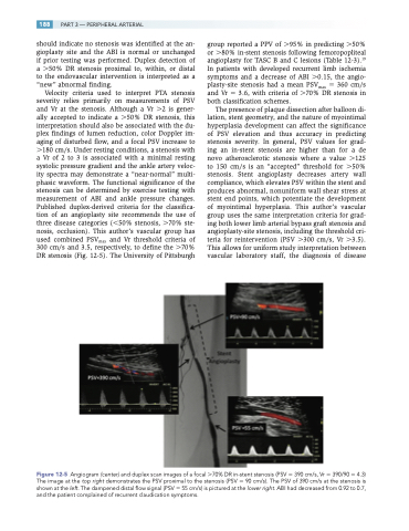
Velocity criteria used to interpret PTA stenosis severity relies primarily on measurements of PSV and Vr at the stenosis. Although a Vr 2 is gener- ally accepted to indicate a 50% DR stenosis, this interpretation should also be associated with the du- plex findings of lumen reduction, color Doppler im- aging of disturbed flow, and a focal PSV increase to 180 cm/s. Under resting conditions, a stenosis with a Vr of 2 to 3 is associated with a minimal resting systolic pressure gradient and the ankle artery veloc- ity spectra may demonstrate a “near-normal” multi- phasic waveform. The functional significance of the stenosis can be determined by exercise testing with measurement of ABI and ankle pressure changes. Published duplex-derived criteria for the classifica- tion of an angioplasty site recommends the use of three disease categories (50% stenosis, 70% ste- nosis, occlusion). This author’s vascular group has used combined PSVmax and Vr threshold criteria of 300 cm/s and 3.5, respectively, to define the 70% DR stenosis (Fig. 12-5). The University of Pittsburgh
group reported a PPV of 95% in predicting 50% or 80% in-stent stenosis following femoropopliteal angioplasty for TASC B and C lesions (Table 12-3).19 In patients with developed recurrent limb ischemia symptoms and a decrease of ABI 0.15, the angio- plasty-site stenosis had a mean PSVmax 360 cm/s and Vr 3.6, with criteria of 70% DR stenosis in both classification schemes.
The presence of plaque dissection after balloon di- lation, stent geometry, and the nature of myointimal hyperplasia development can affect the significance of PSV elevation and thus accuracy in predicting stenosis severity. In general, PSV values for grad- ing an in-stent stenosis are higher than for a de novo atherosclerotic stenosis where a value 125 to 150 cm/s is an “accepted” threshold for 50% stenosis. Stent angioplasty decreases artery wall compliance, which elevates PSV within the stent and produces abnormal, nonuniform wall shear stress at stent end points, which potentiate the development of myointimal hyperplasia. This author’s vascular group uses the same interpretation criteria for grad- ing both lower limb arterial bypass graft stenosis and angioplasty-site stenosis, including the threshold cri- teria for reintervention (PSV 300 cm/s, Vr 3.5). This allows for uniform study interpretation between vascular laboratory staff, the diagnosis of disease
Figure 12-5 Angiogram (center) and duplex scan images of a focal 70% DR in-stent stenosis (PSV 390 cm/s, Vr 390/90 4.3) The image at the top right demonstrates the PSV proximal to the stenosis (PSV 90 cm/s). The PSV of 390 cm/s at the stenosis is shown at the left. The dampened distal flow signal (PSV 55 cm/s) is pictured at the lower right. ABI had decreased from 0.92 to 0.7, and the patient complained of recurrent claudication symptoms.
