
Page 281 - Libro 2
P. 281
17 — Venous Valvular Insufficiency Testing
261
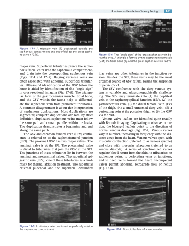
Figure 17-4 A tributary vein ( T ) positioned outside the saphenous compartment and superficial to the great saphe- nous vein (GSV ).
major vein. Superficial tributaries pierce the saphe- nous fascia, enter into the saphenous compartment, and drain into the corresponding saphenous vein (Figs. 17-4 and 17-5). Bulging varicose veins are often associated with abnormal superficial tributar- ies. Ultrasound identification of the GSV below the knee is aided by identification of the “angle sign” in cross-sectional imaging (Fig. 17-6). The triangu- lar form of the gastrocnemius muscle, tibial bone, and the GSV within the fascia help to differenti- ate the saphenous vein from prominent tributaries. A common disagreement is about the interpretation of saphenous duplications. Most duplications are segmental; complete duplications are rare. By strict definition, duplicated saphenous veins must follow the same path and remain parallel within the fascia. The duplication demonstrates a beginning and end along the same path.
The GSV and common femoral vein (CFV) conflu- ence is referred to as the saphenofemoral junction (SFJ). The proximal GSV has two major valves. The terminal valve is at the SFJ. The preterminal valve is distal to tributaries that join the GSV at the SFJ. The junctions of these tributaries lie in between the terminal and preterminal valves. The superficial epi- gastric vein (SEV), one of these tributaries, is a land- mark for thermal ablation treatment. The superficial external pudendal and the superficial circumflex
Figure 17-5 A tributary vein positioned superficially outside the saphenous compartment.
Figure 17-6 The “angle sign” of the great saphenous vein be- low the knee. A triangle is formed by the gastrocnemius muscle (GM), the tibial bone ( T ), and the great saphenous vein (GSV ).
iliac veins are other tributaries in the junction re- gion. Besides the SFJ, these veins may be the most proximal source of GSV reflux, raising the suspicion of pelvic CVVI.
The SSV confluence with the deep venous sys- tem is variable and ultrasonographically challeng- ing. The SSV may terminate into (1) the popliteal vein at the saphenopopliteal junction (SPJ), (2) the gastrocnemius vein, (3) the distal femoral vein (FV) of the thigh, (4) a small unnamed deep vein, (5) a perforating vein at the posterior thigh, or (6) the GSV via the VOG.
Venous valve leaflets are identified quite readily with B-mode imaging. Captivating to observe in mo- tion, the bicuspid leaflets point to the direction of normal venous drainage (Fig. 17-7). Venous valves vary in number, increasing in frequency with the dis- tance away from the heart. Venous valves open with muscular contraction (referred to as venous systole) and close with muscular relaxation (referred to as venous diastole). A series of synchronized valves regulate blood return from the skin, to tributaries, to saphenous veins, to perforating veins or junctions, and to deep veins toward the heart. Incompetent valves permit abnormal retrograde flow or reflux (Fig. 17-8).
Figure 17-7 Bicuspid leaflets of a venous valve.
