
Page 282 - Libro 2
P. 282
262
PART 4 — PERIPHERAL VENOUS
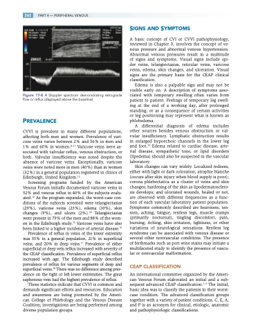
Figure 17-8 A Doppler spectrum demonstrating retrograde flow or reflux (displayed above the baseline).
PREVALENCE
CVVI is prevalent in many different populations, affecting both men and women. Prevalence of vari- cose veins varies between 2% and 56% in men and 1% and 60% in women.8–11 Varicose veins were as- sociated with valvular reflux, venous obstruction, or both. Valvular insufficiency was noted despite the absence of varicose veins. Exceptionally, varicose veins were noted more in men (40%) than in women (32%) in a general population registered in clinics of Edinburgh, United Kingdom.12
Screening programs funded by the American Venous Forum initially documented varicose veins in 32% and venous reflux in 40% of the subjects evalu- ated.13 As the program expanded, the worst-case con- ditions of the subjects screened were telangiectasias (29%), varicose veins (23%), edema (10%), skin changes (9%), and ulcers (2%).14 Telangiectasias were present in 79% of the men and 88% of the wom- en in the Edinburgh study.15 Varicose veins have also been linked to a higher incidence of arterial disease.16
Prevalence of reflux in veins of the lower extremity was 35% in a general population, 21% in superficial veins, and 20% in deep veins.17 Prevalence of either superficial or deep vein reflux increased with severity of the CEAP classification. Prevalence of superficial reflux increased with age. The Edinburgh study described prevalence of reflux for various segments of deep and superficial veins.18 There was no difference among prev- alence on the right or left lower extremities. The great saphenous vein had the highest prevalence of reflux.
These statistics indicate that CVVI is common and demands significant efforts and resources. Education and awareness are being promoted by the Ameri- can College of Phlebology and the Venous Disease Coalition; investigations are being performed among diverse population groups.
SIGNS AND SYMPTOMS
A basic concept of CVI or CVVI pathophysiology, reviewed in Chapter 3, involves the concept of ve- nous pressure and abnormal venous hypertension. Abnormal venous pressures result in a multitude of signs and symptoms. Visual signs include spi- der veins, telangiectasias, reticular veins, varicose veins, edema, skin changes, and ulceration. Visual signs are the primary basis for the CEAP clinical classification.
Edema is also a palpable sign and may not be visible early on. A description of symptoms asso- ciated with temporary swelling often varies from patient to patient. Feelings of temporary leg swell- ing at the end of a working day, after prolonged standing, or as a consequence of certain activities or leg positioning may represent what is known as phlebedema.
A differential diagnosis of edema includes other sources besides venous obstruction or val- vular insufficiency. Lymphatic obstruction results in enlarged hypoechoic channels in the lower leg and foot.19 Edema related to cardiac disease, arte- rial disease, sympathetic tone, or lipid disorders (lipedema) should also be suspected in the vascular laboratory.
Skin changes can vary widely. Localized redness, either with light or dark coloration; atrophie blanche (occurs after skin injury when blood supply is poor); corona phlebectatica as a cluster of veins and skin changes; hardening of the skin as lipodermatosclero- sis develops; and ulcerated wounds, healed or not, are observed with different frequencies as a func- tion of each vascular laboratory patient population. Symptoms commonly described are heaviness, ten- sion, aching, fatigue, restless legs, muscle cramps (primarily nocturnal), tingling discomfort, pain, burning, itching, skin irritation, tightness, or other variations of neurological sensations. Restless leg syndrome can be associated with venous disease or several other nonvascular conditions. The presence of birthmarks such as port wine stains may initiate a multifaceted study to identify the presence of vascu- lar or nonvascular malformation.
CEAP CLASSIFICATION
An international committee organized by the Ameri- can Venous Forum elaborated an initial and a sub- sequent advanced CEAP classification.1–3 The initial, basic idea was to classify the patients in their worst- case condition. The advanced classification groups together with a variety of patient conditions. C, E, A, and P is an acronym for clinical, etiologic, anatomic and pathophysiologic classifications.
