
Page 285 - Libro 2
P. 285
17 — Venous Valvular Insufficiency Testing
265
Figure 17-9 An ultrasound image of the great saphenous vein with a thermal ablation device in place just distal to the saphenofemoral junction (arrow).
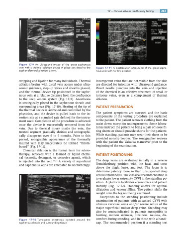
stripping and ligation for many individuals. Thermal ablation begins with distal vein access under ultra- sound guidance, step-up wires and sheaths placed, and the thermal device tip positioned in the saphe- nous vein at a relative distance from the confluence to the deep venous system (Fig. 17-9). Anesthesia is strategically placed in the saphenous sheath and surrounding areas (Fig. 17-10). Heating of the tip of the thermal device is activated and controlled by the physician, and the device is pulled back to the in- sertion site at a standard rate defined for the instru- ment used. Completion of the procedure is achieved once the device is successfully removed from the vein. Due to thermal injury inside the vein, the treated segment gradually shrinks and sonographi- cally disappears over 6 to 9 months. Prior to this period, sonographic appearance of the thermally injured vein may inaccurately be termed “throm- bosed” (Fig. 17-11).
Chemical ablation is the formal term for sclero- therapy, achieved with a foamed or liquid chemi- cal (osmotic, detergent, or corrosive agent), which is injected into the vein.31,32 A variety of superficial and saphenous veins are amenable to sclerotherapy.
Figure 17-10 Tumescent anesthesia injected around the saphenous sheath and surrounding tissue.
Figure 17-11 A postablation ultrasound of the great saphe- nous vein with no flow present.
Incompetent veins that are not visible from the skin are directed for injection with ultrasound guidance. Direct needle puncture into the vein and injection of the chemical is an effective treatment of small or tortuous veins, even as a complement of thermal ablation.
PATIENT PREPARATION
The patient symptoms are assessed and the basic components of the testing procedure are explained to the patient. The patient removes clothing from the waist down except for undergarments. Some labora- tories instruct the patient to bring a pair of loose fit- ting shorts or should provide shorts for the patients. While standing, patients may wear their shoes or be provided nonslip booties. The sonographer reviews with the patient the Valsalva maneuver prior to the beginning of the examination.
PATIENT POSITIONING
The deep veins are evaluated initially in a reverse Trendelenburg position with the head and torso above the thigh, knee, and feet. The focus is to determine patency more so than unsuspected deep venous thrombosis. The classical recommendation is to evaluate lower extremity CVVI in the standing po- sition. A platform facilitates ergonomics and patient stability (Fig. 17-12). Standing allows for optimal dilatation and venous filling. The patient shifts the weight onto the leg not being examined.
Exceptions to the standing positioning include examination of patients with advanced CVVI with obvious varicose veins and/or severe reflux of the major superficial and/or deep veins. Standing may also be contraindicated in patients susceptible to fainting, motion sickness, dizziness, nausea, dis- comfort during standing, and in those with a handi- cap. The recommended position if a standing test
