
Page 287 - Libro 2
P. 287
17 — Venous Valvular Insufficiency Testing
267
Figure 17-14 An adjustable examination stool used by tech- nologists during venous evaluations.
SCANNING TECHNIQUE
The essential protocol includes evaluation of patency and reflux in the deep veins, femoropopliteal veins, particularly the perforating veins, veins in the sa- phenous compartments, nonsaphenous superficial veins, and tributaries. Protocols also include the differential diagnoses of nonvenous pathologies such as arterial and lymphatic abnormalities and masses.
Evaluation of the deep veins precedes any CVVI testing. Detection of acute deep venous thrombo- sis (DVT) is rare; continuation of a CVVI examina- tion becomes secondary and is not recommended. Interpreting and/or referring physicians should be contacted according to preestablished protocols. A preliminary report may be acceptable at certain institutions.
Detection of chronic deep venous obstruction, either total or partial, is part of a CVVI evalua- tion, and the examination is completed. Chronic venous obstruction is suspected in patients with a
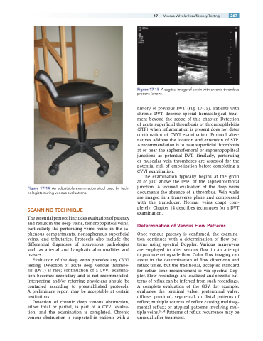
Figure 17-15 A sagittal image of a vein with chronic thrombus present (arrow).
history of previous DVT (Fig. 17-15). Patients with chronic DVT deserve special hematological treat- ment beyond the scope of this chapter. Detection of acute superficial thrombosis or thrombophlebitis (STP) when inflammation is present does not deter continuation of CVVI examination. Protocol alter- natives address the location and extension of STP. A recommendation is to treat superficial thrombosis at or near the saphenofemoral or saphenopopliteal junctions as potential DVT. Similarly, perforating or muscular vein thromboses are assessed for the potential risk of embolization before completing a CVVI examination.
The examination typically begins at the groin at or just above the level of the saphenofemoral junction. A focused evaluation of the deep veins documents the absence of a thrombus. Vein walls are imaged in a transverse plane and compressed with the transducer. Normal veins coapt com- pletely. Chapter 14 describes techniques for a DVT examination.
Determination of Venous Flow Patterns
Once venous patency is confirmed, the examina- tion continues with a determination of flow pat- terns using spectral Doppler. Various maneuvers are employed to alter venous flow in an attempt to produce retrograde flow. Color flow imaging can assist in the determination of flow directions and reflux times, but the traditional, accepted standard for reflux time measurement is via spectral Dop- pler. Flow recordings are localized and specific pat- terns of reflux can be inferred from such recordings. A complete evaluation of the GSV, for example, evaluates the terminal valve; preterminal valve; diffuse, proximal, segmental, or distal patterns of reflux; multiple sources of reflux causing multiseg- mental reflux; or atypical patterns involving mul- tiple veins.33,34 Patterns of reflux recurrence may be unusual after treatment.
