
Page 290 - Libro 2
P. 290
270
PART 4 — PERIPHERAL VENOUS
junction. Transthoracic cardiac US can be used to ob- serve the foam arrival in the right heart.35,36 Transtho- racic cardiac US may show bubbles in the left heart, indicating the presence of a right to left shunt or a patent foramen ovale (PFO). Transcranial Doppler (TCD) US may demonstrate the presence of high in- tensity transient signal (HITS) in the middle cerebral artery during foam sclerotherapy. Not all laboratories perform these adjunctive ultrasounds.
Follow-up US
Postablation protocols include a limited evaluation of the deep veins to ensure patency as well as a com- plete examination of the treated vein. US can demon- strate if segments of the treated vein are fully fibrosed or if the vein is recanalized—totally or partially in diameter as well as completely or segmentally in its longitudinal extension.37,38 A thrombus may be de- tected immediately after treatment or after months or years, due to recanalization and rethrombosis.
Patients are followed primarily because venous disease is constantly reoccurring even in treated extremities. The opposite leg may develop a treat- able disease with time, or the treated leg may have new veins requiring treatment. The patient follow-up study is commonly bilateral and follows the same protocols as described for definitive diagnostic US. A common problem is a lack of adequate history on previous procedures. Technologists often discover that veins are absent or that treated veins are present.
PITFALLS
Equipment settings must be properly adjusted to accurately detect venous reflux. Some technical factors affecting reflux measurement include the following:
• Gain alters the sensitivity of spectral Doppler or color flow
• High persistence may result in false-positive color flow findings
• Velocity scales (physiologic term) or PRF (engi- neering term) also affect the spectral Doppler or color flow sensitivity to detection of reflux
• Different instruments have different settings and different characteristics and may affect reflux detection
There are alternate explanations to retrograde flow, although initially described as “reflux.” Flow from a tributary filling in a segment of the vein after a compression/decompression maneuver may produce a reverse flow pattern if this flow enters below a valve sinus. Valve leakage can occur when valves take a long time to close or if they close but do not remain closed. Valve leakage may be delayed and
occur after several seconds following the testing ma- neuver. Sometimes the valves can close normally, but a symptomatic patient may have valve leakage with every respiration. Reverse flow may occur by surgical correction of hemodynamics to preserve drainage. Flush ligation of the saphenofemoral junction, for example, may create reverse flow through the saphe- nous vein until the next distal perforating vein. This perforating vein thus becomes a treatment-designed junction where flow is shunted or directed to this new drainage point. CHIVA procedures commonly create reverse flow in successfully treated veins in a similar surgical method, as described previously.
DIAGNOSIS
B-MODE ULTRASOUND FINDINGS
Normal B-mode image findings will reveal smooth, thin-walled veins with no obvious change in venous diameter. The vein is fully compressible, and the lumen is hypoechoic.
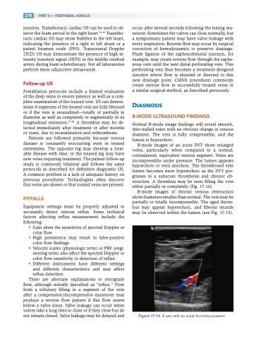
B-mode images of an acute DVT show enlarged veins, particularly when compared to a normal, contralateral, equivalent venous segment. Veins are incompressible under pressure. The lumen appears hypoechoic or even anechoic. The thrombosed vein lumen becomes more hyperechoic as the DVT pro- gresses to a subacute thrombosis and chronic ob- struction. A thrombus may be seen filling the vein either partially or completely (Fig. 17-16).
B-mode images of chronic venous obstruction show diameters smaller than normal. The vein may be partially or totally incompressible. The aged throm- bus may appear hyperechoic, and fibrous strands may be observed within the lumen (see Fig. 17-15).
Figure 17-16 A vein with an acute thrombus present.
