
Page 291 - Libro 2
P. 291
17 — Venous Valvular Insufficiency Testing
271
The veins may display possible tortuosity. Collateral veins develop and enlarge with time.
With chronic venous valvular insufficiency, the vein diameter enlarges, but the veins are completely compressible. The lumen is hypoechoic. Some of the valve sinuses may appear enlarged with flapping valve leaflets. The affected veins eventually become tortuous, varicosed, or even aneurysmal.
Immediately following thermal ablation, the vein is still anesthetized and compressed by tumescence; B-mode imaging may change rapidly or take months to reveal eventual results of treatment. In a postpro- cedural follow-up, typically at 6 to 9 months, the vein may be segmentally sonographically absent, fibrosed, thrombosed, or recanalized simultaneously at different sites along the course of the vein.37,38
SPECTRAL DOPPLER WAVEFORMS
Normal venous flow waveforms are spontaneous, phasic with respiration, and unidirectional toward the heart. Flow augments with distal compression or release of proximal compression (Fig. 17-17).
In the presence of an acute, fully occlusive DVT, the spectral Doppler waveform shows an absence of flow. Partially occlusive DVT, proximal thrombosis, or exter- nal compression can cause continuous flow. The lack of flow augmentation following distal compression or the release of proximal compression is observed in pa- tients with acute DVT. Arterial flow waveforms can be present from within a lysing thrombus.
Flow is also absent with a complete chronic venous obstruction. A partial obstruction, a proxi- mal obstruction, or an external compression can cause continuous flow and lack of flow augmenta- tion, similarly to acute DVT. Spectral analysis may also reveal flow through small, tortuous channels within the diseased vein. Arterial, venous, or fistula- like flow may be observed in small vessels near the obstructed vein, and these may be a possible sign of
Figure 17-17 A normal venous Doppler signal with no reflux present.
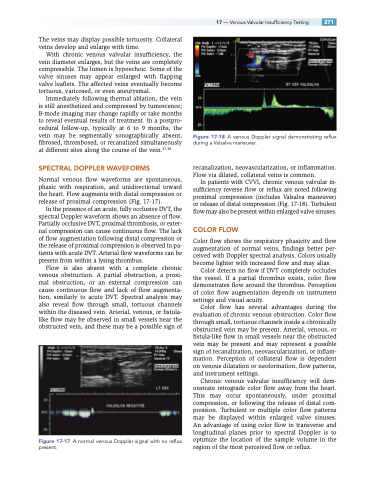
Figure 17-18 A venous Doppler signal demonstrating reflux during a Valsalva maneuver.
recanalization, neovascularization, or inflammation. Flow via dilated, collateral veins is common.
In patients with CVVI, chronic venous valvular in- sufficiency reverse flow or reflux are noted following proximal compression (includes Valsalva maneuver) or release of distal compression (Fig. 17-18). Turbulent flow may also be present within enlarged valve sinuses.
COLOR FLOW
Color flow shows the respiratory phasicity and flow augmentation of normal veins, findings better per- ceived with Doppler spectral analysis. Colors usually become lighter with increased flow and may alias.
Color detects no flow if DVT completely occludes the vessel. If a partial thrombus exists, color flow demonstrates flow around the thrombus. Perception of color flow augmentation depends on instrument settings and visual acuity.
Color flow has several advantages during the evaluation of chronic venous obstruction. Color flow through small, tortuous channels inside a chronically obstructed vein may be present. Arterial, venous, or fistula-like flow in small vessels near the obstructed vein may be present and may represent a possible sign of recanalization, neovascularization, or inflam- mation. Perception of collateral flow is dependent on venous dilatation or neoformation, flow patterns, and instrument settings.
Chronic venous valvular insufficiency will dem- onstrate retrograde color flow away from the heart. This may occur spontaneously, under proximal compression, or following the release of distal com- pression. Turbulent or multiple color flow patterns may be displayed within enlarged valve sinuses. An advantage of using color flow in transverse and longitudinal planes prior to spectral Doppler is to optimize the location of the sample volume in the region of the most perceived flow or reflux.
