
Page 401 - Libro 2
P. 401
24 — Intraoperative Duplex Sonography
381
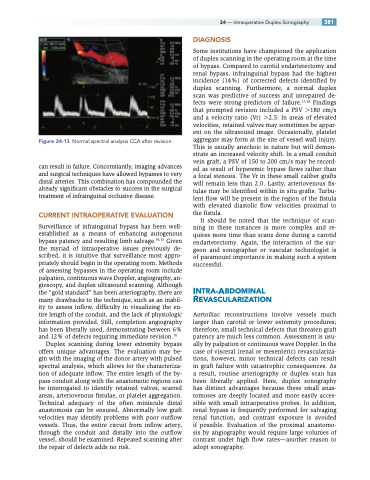
Figure 24-13 Normal spectral analysis CCA after revision.
can result in failure. Concomitantly, imaging advances and surgical techniques have allowed bypasses to very distal arteries. This combination has compounded the already significant obstacles to success in the surgical treatment of infrainguinal occlusive disease.
CURRENT INTRAOPERATIVE EVALUATION
Surveillance of infrainguinal bypass has been well- established as a means of enhancing autogenous bypass patency and resulting limb salvage.14,15 Given the myriad of intraoperative issues previously de- scribed, it is intuitive that surveillance most appro- priately should begin in the operating room. Methods of assessing bypasses in the operating room include palpation, continuous wave Doppler, angiography, an- gioscopy, and duplex ultrasound scanning. Although the “gold standard” has been arteriography, there are many drawbacks to the technique, such as an inabil- ity to assess inflow, difficulty in visualizing the en- tire length of the conduit, and the lack of physiologic information provided. Still, completion angiography has been liberally used, demonstrating between 6% and 12% of defects requiring immediate revision.16
Duplex scanning during lower extremity bypass offers unique advantages. The evaluation may be- gin with the imaging of the donor artery with pulsed spectral analysis, which allows for the characteriza- tion of adequate inflow. The entire length of the by- pass conduit along with the anastomotic regions can be interrogated to identify retained valves, scarred areas, arteriovenous fistulae, or platelet aggregation. Technical adequacy of the often miniscule distal anastomosis can be ensured. Abnormally low graft velocities may identify problems with poor outflow vessels. Thus, the entire circuit from inflow artery, through the conduit and distally into the outflow vessel, should be examined. Repeated scanning after the repair of defects adds no risk.
DIAGNOSIS
Some institutions have championed the application of duplex scanning in the operating room at the time of bypass. Compared to carotid endarterectomy and renal bypass, infrainguinal bypass had the highest incidence (14%) of corrected defects identified by duplex scanning. Furthermore, a normal duplex scan was predictive of success and unrepaired de- fects were strong predictors of failure.17,18 Findings that prompted revision included a PSV 180 cm/s and a velocity ratio (Vr) 2.5. In areas of elevated velocities, retained valves may sometimes be appar- ent on the ultrasound image. Occasionally, platelet aggregate may form at the site of vessel wall injury. This is usually anechoic in nature but will demon- strate an increased velocity shift. In a small conduit vein graft, a PSV of 150 to 200 cm/s may be record- ed as result of hyperemic bypass flows rather than a focal stenosis. The Vr in these small caliber grafts will remain less than 2.0. Lastly, arteriovenous fis- tulae may be identified within in situ grafts. Turbu- lent flow will be present in the region of the fistula with elevated diastolic flow velocities proximal to the fistula.
It should be noted that the technique of scan- ning in these instances is more complex and re- quires more time than scans done during a carotid endarterectomy. Again, the interaction of the sur- geon and sonographer or vascular technologist is of paramount importance in making such a system successful.
INTRA-ABDOMINAL REVASCULARIZATION
Aortoiliac reconstructions involve vessels much larger than carotid or lower extremity procedures; therefore, small technical defects that threaten graft patency are much less common. Assessment is usu- ally by palpation or continuous wave Doppler. In the case of visceral (renal or mesenteric) revasculariza- tions, however, minor technical defects can result in graft failure with catastrophic consequences. As a result, routine arteriography or duplex scan has been liberally applied. Here, duplex sonography has distinct advantages because these small anas- tomoses are deeply located and more easily acces- sible with small intraoperative probes. In addition, renal bypass is frequently performed for salvaging renal function, and contrast exposure is avoided if possible. Evaluation of the proximal anastomo- sis by angiography would require large volumes of contrast under high flow rates—another reason to adopt sonography.
