
Page 399 - Libro 2
P. 399
24 — Intraoperative Duplex Sonography 379
a vascular clamp injury, which may be present and would require attention.
A focal peak systolic velocity (PSV) increase in the internal carotid artery can identify a significant com- plication. Reexamination or revision of the surgical site is warranted if the PSV exceeds 180 cm/s or the internal carotid to common carotid PSV ratio is greater than 2.5. In some patients, this may be associated with a fresh platelet aggregate, which is often unable to be identified on the B-mode image due to its anechoic
PATHOLOGY BOX 24-1
Common Vascular Pathology Observed Intraoperatively
Pathology Observed Ultrasound Characteristics
“Shelf” lesion/ residual lesion
Intimal flap
Dissection
Platelet aggregate
Stenosis: carotid or lower extremity bypass graft
Stenosis: renal or celiac artery
Stenosis: superior mesenteric artery
Arteriovenous fistula
Retained valve
• Hyperechoic plaque project- ing into the vessel lumen
• May display an abrupt edge
• Small projection into the vessel lumen, usually a few millimeter in length
• Disturbed flow or aliasing may be present
• Linear object seen parallel to vessel walls
• Turbulent or disturbed flow present
• Hypoechoic or anechoic material adjacent to vessel wall
• Focal elevation in PSV • Increased Vr
• PSV 180 cm/s
• Vr 2.5
• PSV 200 cm/s
• PSV 275 cm/s
• Patent branch may be seen arising from an in situ bypass
• Turbulence and aliasing pre- sent in area of side branch
• Elevated diastolic velocities
in bypass graft proximal to side branch
• Hyperechoic structure protruding into lumen of vein bypass graft; may be associated with slight dilation of valve sinus
• Turbulence or aliasing may be present
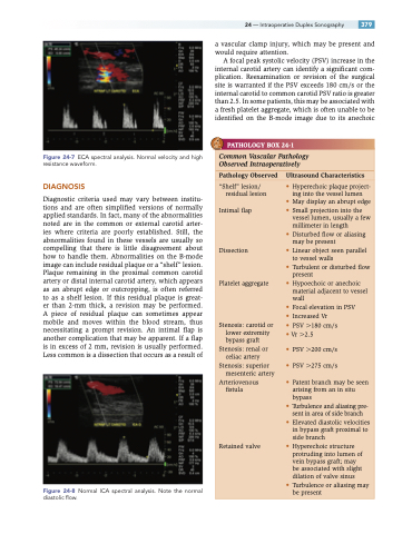
Figure 24-7 ECA spectral analysis. Normal velocity and high resistance waveform.
DIAGNOSIS
Diagnostic criteria used may vary between institu- tions and are often simplified versions of normally applied standards. In fact, many of the abnormalities noted are in the common or external carotid arter- ies where criteria are poorly established. Still, the abnormalities found in these vessels are usually so compelling that there is little disagreement about how to handle them. Abnormalities on the B-mode image can include residual plaque or a “shelf” lesion. Plaque remaining in the proximal common carotid artery or distal internal carotid artery, which appears as an abrupt edge or outcropping, is often referred to as a shelf lesion. If this residual plaque is great- er than 2-mm thick, a revision may be performed. A piece of residual plaque can sometimes appear mobile and moves within the blood stream, thus necessitating a prompt revision. An intimal flap is another complication that may be apparent. If a flap is in excess of 2 mm, revision is usually performed. Less common is a dissection that occurs as a result of
Figure 24-8 Normal ICA spectral analysis. Note the normal diastolic flow.
