
Page 398 - Libro 2
P. 398
378
PART 6 — MISCELLANEOUS
external carotid artery, 6.5% of the collected cases had abnormalities in the internal carotid artery, usually at the distal end of the endarterectomy.1
Routine intraoperative angiography offers the ad- vantage of visualizing the intracranial carotid artery, as well as the cervical area. Lesions proximal in the com- mon carotid artery are usually not assessed, however, and no physiologic data is identified. In an early study using routine completion angiography, Donaldson et al. found 71 defects in a series of 410 carotid endarterec- tomies, warranting correction in 16% of cases. These corrections did not add to morbidity, as the stroke rate remained below 2%.2 Zannetti et al. evaluated 1,305 carotid endarterectomies with completion angiogra- phy in 77% and identified 9% defects, 4% of which were revised. There was an increased stroke rate in this group despite revision. Nevertheless, the overall stroke rate was less than 1%; this raises questions regarding the advantage of routine imaging.3 Westerband et al. reported a 19% incidence of defects requiring repair, with no postoperative occlusions in this group.4
CURRENT INTRAOPERATIVE EVALUATION
Currently, continuous wave Doppler interrogation alone is the most commonly used assessment during carotid endarterectomy. This method has been shown to be quite sensitive, but not specific, identifying ab- normalities in 4.3% in early studies.5 Although this modality is simple and fairly reliable in experienced hands, it usually requires a confirmatory study such as an angiography to justify re-exploration. B-mode ultrasound has also been used in completion stud- ies to determine which findings signal the need for revision. Again, the incidence of complications is so low it is difficult to make recommendations based on these smaller studies.6
Bandyk et al. applied pulse Doppler spectral analy- sis to carotid endarterectomy sites and reported on 250 procedures using this technique.7 In a follow-up study of 461 endarterectomies studied with duplex scanning, less than 6% required intraoperative revision, and the permanent stroke rate was 1.3%. Patients with nor- mal scans had a lower incidence of late postoperative stroke.8 The Mayo Clinic reported results in 87 patients using routine duplex scanning. In the study, 9% had significant findings requiring immediate revision. Stroke rates were 1.9% and were equal between nor- mal and repaired groups. Two of three patients with significant common carotid lesions that were not ad- dressed suffered strokes. These data suggest the safety and efficacy of routine duplex scanning.9 Numerous other small studies have shown similar advantages of intraoperative duplex sonography.10,11
Although completion duplex sonography is intuitive- ly beneficial, caution must be exercised in interpreting
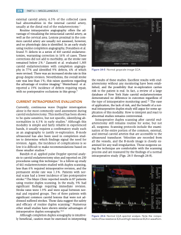
Figure 24-5 Normal grayscale image CCA.
the results of these studies. Excellent results with end- arterectomy without any monitoring have been estab- lished, and the possibility that re-exploration carries risk to the patient is real. In fact, a review of a large database of New York State carotid endarterectomies demonstrated no difference in outcomes regardless of the type of intraoperative monitoring used.12 The ease of application, the lack of risk, and the benefit of a nor- mal intraoperative duplex study still argue for some ap- plication of this modality. How to interpret and react to abnormal studies remains controversial.
Intraoperative duplex scanning after carotid end- arterectomy still remains routine for some, but not all, surgeons. Scanning protocols include the exami- nation of the entire portion of the common, external, and internal carotid arteries that are accessible to the ultrasound transducer. Velocities are recorded from all the vessels, and the B-mode image is closely ex- amined for any wall irregularities. Those surgeons us- ing the technique are comfortable with the scanning process and are reassured by the findings of a normal intraoperative study (Figs. 24-5 through 24-8).
Figure 24-6 Normal CCA spectral analysis. Note the compo- nents of low-resistance ICA and high-resistance ECA in waveform.
