
Page 396 - Libro 2
P. 396
376 PART 6 — MISCELLANEOUS
TABLE 24-1
Common Applications for Intraoperative Vascular Ultrasound
Surgical Procedure
Carotid endarterectomy
Infrainguinal revascularization
Renal and mesenteric artery bypass
SONOGRAPHIC EXAMINATION TECHNIQUES
Vascular reconstructions, which lend themselves to intraoperative application of duplex scanning, in- clude carotid endarterectomy and infrainguinal and visceral bypass. Results of carotid endarterectomy are already consistently excellent, so improvements are likely to be in small increments. Lower extrem- ity bypass results are plagued by problems related to the inflow, outflow, and conduit. Duplex bypass sur- veillance has been shown to enhance patency and limb salvage, and beginning the surveillance in the operating room is a natural extension of that policy. Renal and visceral bypass patency depends on tech- nical excellence, which is easily assessed with du- plex scanning. Table 24-1 summarizes the common vascular applications for intraoperative ultrasound and abnormalities that can be encountered.
SCANNING TECHNIQUE
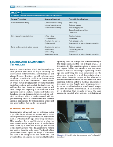
Intraoperative ultrasound can be performed using a portable duplex ultrasound system with a trans- ducer specifically designed for vascular applications such as a “hockey stick” type linear array transducer. This transducer has a small footprint to allow for easy access into the surgical areas. A sterile sheath with a latex tip is filled with sterile gel and used to isolate the transducer while being careful to remove any bubbles from the probe cover. The length of the probe cover allows a significant length of transducer and cord to be brought onto the sterile field. The wound is filled with saline, the overhead lights in the
Anatomy Examined
Common carotid artery Internal carotid artery External carotid artery
Inflow artery Outflow artery Anastomotic regions Entire conduit
Anastomotic regions Renal artery
Celiac artery Mesenteric artery
Potential Complications
Intimal flap
Residual plaque
Platelet aggregate Suture line abnormalities Dissection
Retained valves
AV fistulae
Platelet aggregate
Anastomotic or suture line abnormalities
Residual plaque
Platelet aggregate
Dissection
Anastomotic or suture line abnormalities
operating room are extinguished to make viewing of the image easier, and the scan is begun (Figs. 24-1 through 24-4). The scanning protocol is simple, with the surgeon holding the transducer and the sonog- rapher or vascular technologist optimizing the im- age and controlling the other components on the ultrasound console. In general, long axis imaging is used alone. Grayscale images are first obtained to best visualize small defects not well seen with color scanning. Color is added to facilitate placement of the pulsed spectral Doppler gate. Images and wave- forms are stored in cine loops, as well as still images, to allow for careful interpretation. If an abnormal- ity is identified that prompts revision, the scan process is repeated after revision. In infrainguinal
Figure 24-1
transducer.
Portable color duplex scanner with “hockey stick”
