
Page 397 - Libro 2
P. 397
24 — Intraoperative Duplex Sonography
377
Figure 24-2 Sterile sheath filled with gel, prior to placement of the ultrasound transducer.
revascularization, instillation of papaverine into the bypass is helpful to minimize the effects of vaso- spasm frequently seen in these procedures.
TECHNICAL CONSIDERATIONS
Intraoperative assessment during a vascular recon- struction requires a team that is comfortable with the techniques described as well as the equipment nec- essary for the procedure. Modern color duplex scan- ners are available in many shapes and forms, with many new and easily portable machines enclosed in a simple laptop computer configuration. As noted in the preceding section, specific ultrasound transduc- ers for intraoperative use have been developed for vascular applications in multiple anatomic areas. Operating rooms with extensive experience with this technology frequently have dedicated ultrasound machines kept in the operating room at all times.
Figure 24-3 Ultrasound transducer brought onto the surgical field.
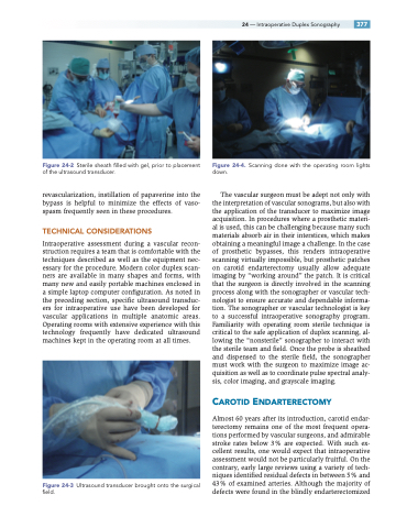
Figure 24-4. Scanning done with the operating room lights down.
The vascular surgeon must be adept not only with the interpretation of vascular sonograms, but also with the application of the transducer to maximize image acquisition. In procedures where a prosthetic materi- al is used, this can be challenging because many such materials absorb air in their interstices, which makes obtaining a meaningful image a challenge. In the case of prosthetic bypasses, this renders intraoperative scanning virtually impossible, but prosthetic patches on carotid endarterectomy usually allow adequate imaging by “working around” the patch. It is critical that the surgeon is directly involved in the scanning process along with the sonographer or vascular tech- nologist to ensure accurate and dependable informa- tion. The sonographer or vascular technologist is key to a successful intraoperative sonography program. Familiarity with operating room sterile technique is critical to the safe application of duplex scanning, al- lowing the “nonsterile” sonographer to interact with the sterile team and field. Once the probe is sheathed and dispensed to the sterile field, the sonographer must work with the surgeon to maximize image ac- quisition as well as to coordinate pulse spectral analy- sis, color imaging, and grayscale imaging.
CAROTID ENDARTERECTOMY
Almost 60 years after its introduction, carotid endar- terectomy remains one of the most frequent opera- tions performed by vascular surgeons, and admirable stroke rates below 3% are expected. With such ex- cellent results, one would expect that intraoperative assessment would not be particularly fruitful. On the contrary, early large reviews using a variety of tech- niques identified residual defects in between 5% and 43% of examined arteries. Although the majority of defects were found in the blindly endarterectomized
