
Page 43 - Libro 2
P. 43
2 — Arterial Physiology
23
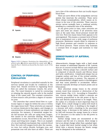
Volume retention
just a few of the substances that can locally impact blood flow.
There are nerve fibers of the sympathetic nervous system that innervate the arterioles. These nerve fibers release norepinephrine, which causes an in- crease in the tone of the arterioles. These vasocon- strictor nerves normally have a continual activity, resulting in a contractile tone of the arterioles.
At any given point in time, some arterioles are open and some are closed. If all arterioles were open at the same time, blood pressure would fall very low. Flow into many tissue beds appears to be autoregulated. This means a constant level of blood flow is maintained over a wide range of perfusion pressures. Resistance vessels dilate in response to high blood pressure and constrict in response to low blood pressure. These actions help maintain a constant flow of oxygen and nutrients to vital organs.
HEMODYNAMICS OF ARTERIAL DISEASE
Atherosclerotic changes begin with a lipid streak that consists of subintimal deposits of fat. Lesions that are of concern include fibrous and complicated plaques. Fibrous plaque has a smooth surface and is composed of smooth muscle and fibrous tissue and lacks calcification. Complicated plaque has an irregular surface and loss of the normal endothe- lium and calcification is present. The exposure of the subendothelial collagen matrix is thrombogenic and may cause platelets to accumulate. Athero- sclerosis typically develops at branch points and at bifurcations.
Most abnormal energy losses in the arterial system result from stenoses or obstruction of the vessel lumen (Fig. 2-11). According to Poiseuille’s law, viscous energy losses within a stenosis are inversely proportional to the 4th power of the ra- dius and directly proportional to its length. Thus, the radius of a stenosis is more important than its length. Even a small change in radius will result in large changes in flow. A doubling in the length of a stenosis will yield a doubling in the associated energy losses. A decrease in the radius of a vessel by half will increase the energy losses by a factor of 16 (because it is the radius to the 4th power in Poiseuille’s law).
Inertial energy losses are encountered at the entrance and the exit of a stenosis. More energy is lost at an abrupt change rather than a gradual taper- ing. A great deal more inertial energy is lost when blood exits a stenosis because the kinetic energy may be dissipated in the turbulent jet.
A Systole
Volume release
B Diastole
Figure 2-10 A diagram illustrating the distensibility of the arterial walls, (A) systole expanding the vessel walls, (B) an- tegrade flow in diastole produced by the elastic recoil of the vessels.
CONTROL OF PERIPHERAL CIRCULATION
Peripheral circulation is controlled centrally by the nervous system and locally by conditions at the tissue bed. Vessels involved in regulating blood flow are called the resistance vessels, the arteri- oles. The vessel diameter is varied by contracting or relaxing the smooth muscle cells in the medial layer of the vessel wall. The constant contraction of these muscle cells provides a degree of vasomo- tor tone.
The arterioles that control blood flow to a par- ticular region or organ lie within the area or within the organ tissue itself. These arterioles are exposed to various chemicals in that region, and changes in concentrations of many substances can impact the arterioles. For instance, if interstitial oxygen lev- els fall because the cells are using more oxygen, this results in the arterioles dilating. When this vasodilation occurs, blood flow to the area will in- crease and bring more oxygen. This is an example of a local feedback mechanism that controls blood flow. Not only will oxygen levels alter the vasomo- tor tone, but carbon dioxide, hydrogen ions, and potassium ions will also have an effect. These are
