
Page 73 - Libro 2
P. 73
4 — The Extracranial Duplex Ultrasound Examination
53
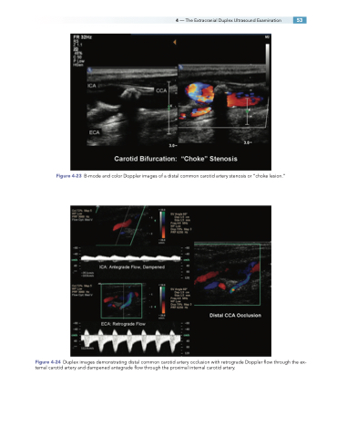
Figure 4-23 B-mode and color Doppler images of a distal common carotid artery stenosis or “choke lesion.”
Figure 4-24 Duplex images demonstrating distal common carotid artery occlusion with retrograde Doppler flow through the ex- ternal carotid artery and dampened antegrade flow through the proximal internal carotid artery.
