
Page 75 - Libro 2
P. 75
4 — The Extracranial Duplex Ultrasound Examination
55
Figure 4-26 Doppler flow signal obtained in the common ca- rotid artery, with arrhythmia.
Differentiating low cardiac output from aortic steno- sis can be difficult with duplex ultrasound. However, an unusual waveform with two prominent systolic peaks separated by a systolic retraction called “pul- sus bisferiens” has been described in the carotid arteries of patient with aortic valvular disease and hypertrophic obstructive cardiomyopathy.11 Cardiac arrhythmias (abnormal heart rhythms or rates) can also make interpreting Doppler waveforms difficult because standard velocity criteria may not apply and the waveform contour may be altered (Fig. 4-26).
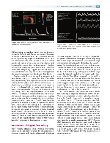
Cardiac assist devices are used in patients with heart failure to support cardiac function, often while they are recovering from myocardial infarction or heart surgery. Some cardiac assist devices are intended for short-term use, whereas others may be in place for longer periods as a bridge to cardiac transplantation. A ventricular assist device (VAD) and an intra-aortic bal- loon pump (IABP) are two examples of cardiac assist devices. The effects of cardiac assist devices on Dop- pler arterial waveform contour is profound, creating patterns that may be unrecognizable as arterial flow. An example of a carotid artery Doppler waveform in a patient with an IABP is shown in Figure 4-27. There- fore, a “disclaimer” is necessary in the vascular labo- ratory report stating that waveform contour cannot be interpreted according to standard criteria. Hospital- based vascular laboratories are most likely to encoun- ter these patients, as they are almost always inpatients. However, some of these devices are portable, so oc- casionally an outpatient may present to the vascular laboratory with one of these devices in place.
Measurement of Doppler Flow Velocity
Flow velocity obtained from Doppler waveforms serves as the primary criterion for classification of stenosis severity with duplex ultrasound. Obtaining
Figure 4-27 Carotid artery Doppler flow affected by cardiac assist devices: intra-aortic balloon pump (IABP) and left ven- tricular assist device (LVAD).
accurate Doppler information is highly dependent on proper examination technique, particularly using the correct angle of insonation. The Doppler angle of insonation is traditionally defined as the angle be- tween the line of the ultrasound beam and the arteri- al wall at the site of the PW Doppler sample volume. This assumes that the direction of flow is parallel to the wall of the artery and requires that the angle cursor be aligned parallel to the vessel wall. How- ever, “off-axis” flow (flow not parallel to the wall) is common in diseased arteries. When the direction of an off-axis flow jet can be determined based on the color Doppler image, some laboratories set the angle cursor parallel to the flow jet, but it has not been es- tablished that this is more accurate than setting the angle cursor parallel to the vessel wall.
All arterial velocity measurements should be ob- tained using an angle of insonation of 60° or less. This is accomplished by either adjusting the steer- ing of the Doppler beam or by a transducer maneu- ver called “toe-heel.” This maneuver involves slight transducer pressure at either end of the transducer (the toe or the heel) to push a vessel into a slight angle. This moves the vessel enough to create an angle of 60° or less. Although an in-depth discus- sion of Doppler physics is beyond the scope of this chapter, this recommendation is based on the con- cept that errors related to Doppler angles are more critical at large angles because the cosine changes more rapidly at angles approaching 90°.12 Doppler shift frequencies also become very small at larger angles, which further reduces the accuracy of ve- locity measurements. For carotid duplex scanning, a Doppler angle of 60° or less results in clinically valid velocity information for the classification of stenosis severity.
