
Page 76 - Libro 2
P. 76
56 PART 2 — CEREBROVASCULAR
Obtaining the most complete and accurate rep- resentation of flow velocity changes throughout the carotid arteries requires that the PW Doppler sample volume be moved slowly and continuously (“swept”) along the vessel. Simply “spot checking” the flow pattern may overlook very focal or localized flow disturbances. In a segment where stenosis is suspected, sweep the sample volume cursor through the area, proximal to distal, evaluating flow at close- ly spaced intervals and moving the sample volume across the lumen to identify the highest velocity flow jet. As this is being done, the Doppler angle must be continually evaluated and adjusted to maintain proper alignment through each segment.
The carotid and vertebral artery systems are con- nected by the circle of Willis at the base of brain. The branches of the circle of Willis are variable and pro- vide numerous collateral pathways to compensate for extracranial cerebrovascular disease. Potential collateral routes include posterior-to-anterior, side- to-side, and extracranial-to-intracranial. When there is a severe stenosis or complete occlusion of one ICA, velocities may be increased in the contralateral carotid system due to compensatory collateral flow. It is important to recognize this situation to avoid overestimating the extent of disease on the contralat- eral side. Compensatory flow is generally associated with a diffuse increase in flow velocity throughout the contralateral CCA and ICA, without a focal high- velocity jet or other localized flow disturbance. Even when a hemodynamically significant ICA stenosis is identified contralateral to a severe stenosis or
TABLE 4-2
occlusion, it is appropriate to comment that the Dop- pler flow velocity elevation may be due, in part, to the contralateral disease. More detailed information on specific intracranial collateral pathways can be obtained by a transcranial Doppler examination, as discussed in Chapter 7.
CRITERIA FOR CLASSIFICATION OF DISEASE
The B-mode imaging and Doppler waveform param- eters for classification of carotid artery disease have been developed by comparing the results of duplex scanning with “gold standard” imaging modalities or surgical findings. These alternative standard imaging approaches include catheter contrast arteriography, CTA, and magnetic resonance arteriography (MRA). The majority of duplex ultrasound carotid criteria have been validated for the ICA. Therefore, it must be emphasized that such criteria cannot be applied to the CCA or ECA.
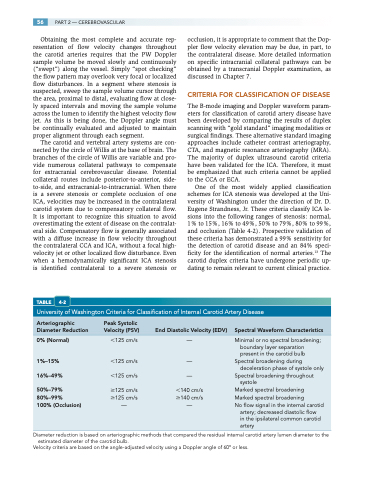
One of the most widely applied classification schemes for ICA stenosis was developed at the Uni- versity of Washington under the direction of Dr. D. Eugene Strandness, Jr. These criteria classify ICA le- sions into the following ranges of stenosis: normal, 1% to 15%, 16% to 49%, 50% to 79%, 80% to 99%, and occlusion (Table 4-2). Prospective validation of these criteria has demonstrated a 99% sensitivity for the detection of carotid disease and an 84% speci- ficity for the identification of normal arteries.13 The carotid duplex criteria have undergone periodic up- dating to remain relevant to current clinical practice.
University of Washington Criteria for Classification of Internal Carotid Artery Disease
Arteriographic Peak Systolic
Diameter Reduction Velocity (PSV) End Diastolic Velocity (EDV) Spectral Waveform Characteristics
0% (Normal)
1%–15%
16%–49%
50%–79% 80%–99%
100% (Occlusion)
125 cm/s 125 cm/s
125 cm/s
125 cm/s 125 cm/s —
—
—
— 140 cm/s
140 cm/s —
Minimal or no spectral broadening; boundary layer separation present in the carotid bulb
Spectral broadening during deceleration phase of systole only
Spectral broadening throughout systole
Marked spectral broadening Marked spectral broadening
No flow signal in the internal carotid
artery; decreased diastolic flow in the ipsilateral common carotid artery
Diameter reduction is based on arteriographic methods that compared the residual internal carotid artery lumen diameter to the estimated diameter of the carotid bulb.
Velocity criteria are based on the angle-adjusted velocity using a Doppler angle of 60° or less.
