
Page 102 - Libro vascular I
P. 102
Chap-08.qxd 1~9~04 16:41 Page 93
ULTRASOUND ASSESSMENT OF THE EXTRACRANIAL CEREBRAL CIRCULATION
93
substantial areas of atheroma can be visualized, and this is most likely to occur at the carotid bifurca- tion. However, in a small proportion of patients, significant disease may be seen in the CCA and may even involve the CCA origin. It is important to remember the way ultrasound interacts with tis- sue and the effects of scanner setup, such as gain control and compression curve selection (Ch. 2), before drawing conclusions about the appearance of plaque surface or composition. A high-frequency transducer should be used when investigating plaque composition. There have been many stud- ies carried out comparing the ultrasound appear- ance of atheromatous plaque with histological investigation of specimens removed during carotid endarterectomy (Fig. 8.13) in an attempt to pre- dict which plaques are more likely to be the source of emboli. Several of these studies show an associ- ation between the symptoms and the presence of intraplaque hemorrhage (i.e., bleeding into the plaque) (Merrit & Bluth 1992). If the surface of a plaque containing intraplaque hemorrhage or lipid pools ruptures, the contents of the plaque are dis- charged into the vessel lumen, causing distal embolization and leading to symptoms such as TIA or stroke. A multicenter European study (European Carotid Plaque Study Group 1995) showed that the echogenicity on the B-mode image was inversely related to the content of soft tissue (including hemorrhage or lipid) and directly related to the presence of calcification. In this study, they described the plaque by using a scale of 1 to 3, with 1 repre- senting ‘strong’ echoes and 3 representing low echogenicity or anechoic areas. The plaques were also described as being homogeneous or hetero- geneous. Irregularity of the plaque surface was not found to relate well to the presence of ulceration.
An international consensus meeting (de Bray et al 1997) used a similar method of describing plaque features: echogenicity (from anechoic to hyperechoic), surface (from smooth to cavitated) and texture (from homogeneous to heterogeneous). It was suggested that echogenicity can be stan- dardized against blood (anechoic), mastoid muscle (isoechogenic) or bone (hyperechogenic cervical vertebrae). Lumen surface was classified as regular, irregular (0.4–2 mm) and ulcerated (2 mm depth and 2 mm in length with well-defined back
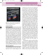
V A
Figure 8.12 Color flow image of the vertebral artery (A) and vein (V) seen between the vertebral processes of the spine (marked by the arrows).
B-MODE IMAGING
Normal appearance
The normal vessel walls will often appear as a double-layer structure when imaged in longitudi- nal section (Fig. 8.7), especially if a high-frequency transducer is used. This represents the intima- media layer and adventitia (Ch. 5) and is most clearly seen on the posterior wall in the CCA, when the vessel lies at right angles to the ultrasound beam. The normal thickness of the intima-media layer is of the order of 0.5 mm (Pignoli et al 1986). A normal vessel lumen should appear anechoic, but it is possible for the sonographer to remove echoes from within the lumen by reducing the time gain compensation (Ch. 2), so careful use of the imaging controls is important. Reverberation artifacts can also give the appearance of structures within the lumen. Occasionally, it is difficult to obtain adequate B-mode images of the bifurcation. In this case, color flow imaging may help locate the vessels and enable spectral Doppler measurements to be made.
Abnormal appearance
The ultrasound appearance of the early stages of carotid artery disease is a thickening of the intima- media layer. As the disease progresses, more
