
Page 101 - Libro vascular I
P. 101
Chap-08.qxd 1~9~04 16:41 Page 92
92
PERIPHERAL VASCULAR ULTRASOUND
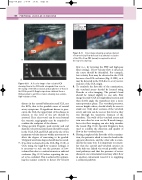
Figure 8.10
ICA
ECA
CCA
WF Low
PRF 1500 Hz Flow Opt: Med V
A
B
the stump of the ICA occlusion and an absence of flow in the ICA beyond. B: Doppler spectrum obtained from a CCA proximal to an ICA occlusion showing low-volume, high-resistance flow.
disease in the carotid bifurcation and ICA, not the ECA, that is the possible cause of carotid artery symptoms. If significant disease is pres- ent in the ICA, the upper limit of the disease in relation to the level of the jaw should be assessed. If no clear vessel can be seen beyond the stenosis, angiography may be required to confirm the endpoint of the disease.
6. Using spectral Doppler, peak systolic and end diastolic velocity measurements should be made in the CCA, ICA and ECA and at the site of the maximum velocity increase within any stenoses to allow the degree of narrowing to be graded. Untypical waveform shapes should also be noted.
7. If no flow is detected in the ICA (Fig. 8.10) or CCA using the high-flow scanner settings, it is necessary to rule out the presence of low- volume flow due to a critical stenosis or subtotal occlusion (Fig. 8.11) before reporting the ves- sel to be occluded. This is achieved by optimiz- ing the scanner controls to detect low-velocity
Figure 8.11 Color image showing a narrow channel of low-velocity flow detected in a subtotal occlusion of the ICA. A low PRF (arrow) is required to detect the low-velocity flow.
flow (i.e., by lowering the PRF and high-pass filter setting). If low-velocity flow is detected, the cause should be identified. For example, low-velocity flow may be detected in the CCA because of an ICA occlusion (Fig. 8.10B), or it may be detected in the ICA due to a very severe stenosis of the ICA origin.
8. To conclude the first side of the examination, the vertebral artery should be located using B-mode or color imaging. The patient’s head should be turned slightly to one side. First image the mid-CCA in longitudinal section and then slowly angle the transducer into a more anteroposterior plane. The vertebral processes, seen as bright echoes, should slowly be seen to stand out. Only short sections of the vertebral artery and vein can be seen at this level as they run through the transverse foramen of the vertebrae. The walls of the vertebral artery and vein can often be seen on the B-mode image, but color flow imaging can also help visualize the vessels (Fig. 8.12). Spectral Doppler is then used to confirm the direction and quality of flow in the vertebral artery.
9. Having completed the first side of the examina- tion, the patient is asked to turn the head in the opposite direction, and the other side is exam- ined in the same way. It is important to remem- ber that the carotid and vertebral arteries on both sides are linked via several possible colla- teral pathways and that the presence of severe disease in one extracranial vessel may affect flow in another extracranial vessel if it is supplying a collateral pathway.
A: A color image of an occluded ICA showing flow in the CCA with retrograde flow seen in
