
Page 121 - Libro vascular I
P. 121
Chap-09.qxd 29~8~04 14:46 Page 112
112
PERIPHERAL VASCULAR ULTRASOUND
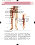
Lumbar artery
Deep iliac circumflex artery
Common femoral artery
Lateral circumflex artery Profunda femoris artery
Superficial femoral artery
Descending genicular artery
Renal artery Aorta
Common iliac artery Internal iliac artery
Inferior epigastric artery Superficial epigastric artery
Medial circumflex artery
Popliteal artery
Sural artery Inferior medial genicular artery
Adductor canal
Above-knee popliteal artery
Superior lateral genicular artery
Inferior lateral genicular artery
Recurrent anterior tibial Proximal anterior
artery
Anterior tibial artery
Dorsalis pedis artery
Tibioperoneal trunk
Posterior tibial artery
tibial artery Peroneal artery
AB
Figure 9.1 A: Arterial anatomy of the aortoiliac and lower limb arteries from an anterior view. B: Arterial anatomy of the lower limb from a posterior view.
aortic bifurcation. The CIA is variable in length (3.5–12 cm) and in some cases it is very short, with the iliac bifurcation occurring close to the aorta. The CIA divides into the external and internal iliac arteries at the iliac bifurcation, which lies deep in the pelvis. The internal iliac artery supplies blood to the pelvis and pelvic viscera. The external iliac artery varies in length (6–12cm) and gives off the deep circumflex iliac artery and inferior epigastric artery, before becoming the common femoral artery (CFA)
at the level of the inguinal ligament. The aorta and iliac arteries lie behind the peritoneum, containing the bowel, which can make imaging of these vessels difficult due to overlying bowel gas. One branch of the CFA that can often be identified with ultrasound is the superficial epigastric artery.
The CFA divides into the deep femoral artery, also known as the profunda femoris artery, and the superficial femoral artery (SFA) at the level of the groin. The profunda femoris artery usually runs
