
Page 123 - Libro vascular I
P. 123
Chap-09.qxd 29~8~04 14:46 Page 114
114
PERIPHERAL VASCULAR ULTRASOUND
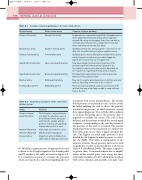
Table 9.1 Common collateral pathways of the lower limb arteries
Diseased artery
Common iliac artery
External iliac artery Common femoral artery
Superficial femoral artery
Superficial femoral artery Popliteal artery
Proximal tibial arteries
Distal normal artery
External iliac artery
Common femoral artery Femoral bifurcation
Above-knee popliteal artery
Below-knee popliteal artery Distal popliteal artery Distal tibial arteries
Common collateral pathway
Lumbar arteries communicating with the iliolumbar arteries of the ipsilateral internal iliac artery, which supply the external iliac artery via retrograde flow; there can also be communication between the contralateral internal iliac artery and ipsilateral internal iliac artery
Ipsilateral internal iliac artery via pelvic connections to the deep iliac circumflex artery or inferior epigastric artery
Ipsilateral pelvic arteries filling the profunda femoris artery via the femoral circumflex arteries, which supply the superficial femoral artery via retrograde flow
Flow via profunda femoris artery (or branches of the proximal superficial femoral artery if patent) to the descending or superior genicular arteries, depending on the length of the superficial femoral artery occlusion
Profunda femoris artery branches to inferior genicular branches of the popliteal artery
Flow via the superior genicular arteries to inferior genicular arteries, depending on the level of the occlusion
There are numerous arterial collateral connections in the calf, but they may not be large enough to carry sufficient flow to the foot
Table 9.2 Anatomical variations of the lower limb arterial system
Artery
Common femoral artery bifurcation
Anterior tibial artery Anterior tibial artery Peroneal artery
Variation
The bifurcation can sometimes be very high; the proximal course of the profunda femoris artery can sometimes be variable and lies posterior medial to the superficial femoral artery in 5% of cases
High origin across the knee joint May be small or hypoplastic
Origin from anterior tibial artery rather than the tibioperoneal trunk
in patients with severe arterial disease. The systolic blood pressure is measured at each of these points by briskly inflating the cuff to above the patient’s systolic blood pressure, at which point the arterial flow signal disappears. The cuff should be inflated to at least 30mmHg above the pressure that is required to occlude the artery. The cuff is then deflated, and the pressure at which the arterial signal reappears, corresponding to the systolic pressure at the position of the cuff, is recorded. The systolic brachial pressure is then measured in a similar way from both arms, in case there is upper extremity dis- ease. The highest recorded ankle pressure is then divided by the highest brachial pressure to calcu- late the ABPI. This index is independent of the patient’s systemic blood pressure and can be used to grade the severity of arterial disease as shown in Table 9.3 (AbuRahma 2000). The index is equal to, or greater than, 1 in normal subjects due to amplifi- cation of the arterial pulse wave along the limb. Conversely, an index of 0.25 would indicate a patient with severe ischemia and possible rest pain. Care
(8–10 MHz) continuous wave Doppler probe is used to listen to the Doppler signals in the dorsalis pedis and PT arteries at the ankle, as shown in Figure 9.3. It is sometimes necessary to examine the peroneal artery, as it may be the only vessel supplying the foot
