
Page 155 - Libro vascular I
P. 155
Chap-11.qxd 29~8~04 14:49 Page 146
146
PERIPHERAL VASCULAR ULTRASOUND
be dependent on factors such as physical size, sex and age.
ANATOMY OF THE ABDOMINAL AORTA
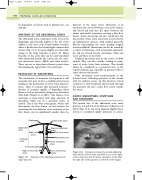
The abdominal aorta commences at the level of the diaphragm and descends slightly to the left of the midline to the level of the fourth lumbar vertebra, where it divides into the left and right common iliac arteries (Fig. 11.1). It tapers slightly as it descends, owing to the large branches it gives off. Major branches of the aorta that can be easily identified with ultrasound include the coeliac axis, the supe- rior mesenteric artery (SMA) and renal arteries. These can act as important reference points when determining the upper limit of an aneurysm.
PATHOLOGY OF ANEURYSMS
The mechanism of aneurysm development is still uncertain but may involve a multifactorial process leading to the destruction of aortic wall connective tissue. There is evidence that increased local pro- duction of enzymes capable of degrading elastic fibers as well as interstitial collagens are associated with AAA (Wassef et al 2001). The lumen of an aneurysm is often lined with large amounts of thrombus, which can be a potential source of emboli. This is also why arteriograms, which only demonstrate the flow lumen, are not accurate for estimating the true diameter of an aneurysm, as the flow lumen can be significantly smaller than the
diameter of the entire vessel. Dissection of an aneurysm can occur following a tear in the intima, and blood can leak into the space between the intima and media, sometimes creating a false flow lumen. Aortic aneurysms can also extend into the iliac arteries. Some aortic aneurysms are involved in an inflammatory process, with marked periaortic fibrosis surrounding the aorta making surgical resection difficult. Aneurysms can also be caused by a variety of infections, such as bacterial endocardi- tis, and are termed mycotic aneurysms. These can occur anywhere in the body.
Popliteal aneurysms may be the source of distal emboli. They can also occlude, leading to symp- toms of acute lower limb ischemia. This should always be considered as a potential cause of the acutely ischemic leg, especially in patients with no other obvious risk factors.
False aneurysms occur predominantly in the femoral artery following puncture of the arterial wall for catheter access. In this situation, blood continues to flow backward and forward through the puncture site into a false flow cavity outside the artery.
AORTIC ANEURYSMS: SYMPTOMS AND TREATMENT
The normal size of the abdominal aorta varies between 1.4 and 2.5 cm in diameter (Johnston et al 1991) (Fig. 11.2). An aortic diameter slightly above 2.5cm is considered mildly abnormal or ectatic.
Diaphragm
Right renal artery
Infrarenal aorta
Aortic bifurcation
Right common iliac artery
Celiac trunk (axis)
Superior mesenteric artery
Left renal artery
Abdominal aorta
Inferior mesenteric artery
Left common iliac artery
Suprarenal aorta
IVC
A
Figure 11.1 Anatomy of the abdominal aorta and its major branches.
A transverse image of a normal abdominal aorta (A). The inferior vena cava (IVC) is seen to the right of the aorta (note that the probe orientation means that
the right side of the patient is on the left of the image).
Figure 11.2
