
Page 157 - Libro vascular I
P. 157
Chap-11.qxd 29~8~04 14:49 Page 148
148
PERIPHERAL VASCULAR ULTRASOUND
the graft. Postoperatively, patients normally spend a day or two in intensive care and usually leave hospital 10–14 days after surgery. The elective mor- tality rate for open repair is in the region of 5% (Akkersdijk et al 1994). However, surgically unfit patients have a risk of much higher morbidity and mortality rates.
Endovascular repair
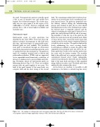
Endovascular repair of aortic aneurysms was described in the early 1990s. There have been sig- nificant technical developments in this field since that time, and several types of commercially manu- factured grafts are now available. The prosthetic stent graft is introduced through an arteriotomy made in the femoral artery and deployed in the aorta to exclude flow into the aneurysm sac. The grafts are made of a synthetic material such as dacron and polytetrafluoroethylene (PTFE) and are supported on an expandable metal framework, or skeleton, of nitonol or stainless steel to prevent kinks and twist- ing. Nowadays, almost all endovascular grafts are bifurcating devices (Fig. 11.3). These are modular systems with the graft supplied in two parts. The bulk of the graft consists of the main body, one complete limb and the short stump of the second
limb. The remaining modular limb is delivered sep- arately via an arteriotomy in the contralateral com- mon femoral artery. The grafts are prepacked onto the delivery catheter during the manufacturing process and retained in place by an outer sheath until deployment in the aorta. During the procedure the femoral artery is surgically exposed, and the catheter containing the main graft is inserted over a guide wire and positioned with the aid of an imag- ing intensifier so that the top of the graft lies just below the renal arteries in the proximal neck. Many of these devices have uncovered metal stents that extend across the renal arteries (suprarenal fixation) to hold the device in place. The graft is deployed by slowly withdrawing the outer covering sheath. A soft balloon is inflated to ensure the graft is fully expanded in the proximal neck, just above the sac. Some grafts have hooks at the top that anchor into the aortic wall for further security. The modular limb is then delivered on a separate catheter via the contralateral femoral artery. Under radiographic control it is positioned so that it fits into the stunted limb of the main body and then is fully expanded using a balloon to make a seal. The distal end is then anchored in the common iliac artery.
As the devices are modular, it is possible to add extensions to the limbs to exclude long iliac artery
Figure 11.3 An example of endovascular aortic aneurysm repair. Note that the left limb of the device is delivered on a separate catheter. Image supplied by courtesy of W L Gore & Associates (UK) Ltd.
