
Page 159 - Libro vascular I
P. 159
Chap-11.qxd 29~8~04 14:49 Page 150
150
PERIPHERAL VASCULAR ULTRASOUND
size of the aneurysm on a serial basis. A screening scan can be performed in 5 to 10min, but more detailed scans or follow-up of endovascular grafts may take 20–30 min.
No special preparation is required, although some units use a bowel preparation to improve visu- alization of the aorta; however, for screening scans this is rarely necessary. The patient should be lying supine with the head supported on a pillow and the arms resting by the sides. Sometimes the patient may have to roll to one side to improve visualiza- tion. The scanner should be configured for an aor- tic investigation but, in the absence of a specific preset, a general abdominal examination setup should be selected. Ensure that the image depth set- ting is not too shallow or too deep. A depth setting of 10–12cm is usually sufficient for the average- sized patient. A 3.5MHz curved linear array trans- ducer, or broad-band equivalent, is the most suitable probe for this investigation. Harmonic imaging can be useful for improving the image quality. In very obese patients a 2–4MHz phased array transducer can help to identify the aorta.
SCANNING TECHNIQUE
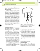
The following description is for a comprehensive investigation of the aorta. However, some depart- ments only perform screening scans and the maxi- mum diameter of the aorta is the only measurement required. Measurements of diameter should be made from a number of different positions. In addi- tion, the shape of the aneurysm and features such as tortuosity or dissection should be documented. The scanning technique for imaging the aorta is demon- strated in Figure 11.5. The procedure is as follows:
1. The aorta is usually easiest to identify by start- ing with the transducer in a transverse image plane, approximately 3–4 cm above the umbili- cus. The aorta is then imaged throughout its visible length, from the upper abdomen above the celiac axis, or SMA, to the aortic bifurca- tion. Where appropriate, the level of the renal arteries can be identified using color flow imag- ing as described later in this chapter. However, it is frequently impossible to image these vessels in the presence of a large aneurysm.
Aorta
B A
C
Transducer positions for scanning the abdominal aorta. A: Transverse. B: Sagittal or longitudinal. C: Coronal. The coronal view is used for measuring the lateral diameter of the aorta (i.e., side to side).
Figure 11.5
2. The abdominal aorta is then imaged in a longi- tudinal or sagittal plane, from the midline along its length to the aortic bifurcation.
3. The aorta is then viewed from a coronal scan plane throughout its length in a longitudinal view to obtain more accurate measurements of the lateral diameter of the aorta (side to side).
4. It is good practice to assess the proximal iliac arteries in transverse and longitudinal scan planes (see Ch. 9) to exclude an isolated iliac artery aneurysm or to define the lower limit of an aneurysm if it extends into the iliac arteries (see Fig. 11.16).
ULTRASOUND APPEARANCE
Normal appearance
The aorta should measure less than 2.5cm at its maximum diameter (Fig. 11.2) and there is usually slight tapering of the aorta from top to bottom. In
