
Page 160 - Libro vascular I
P. 160
Chap-11.qxd 29~8~04 14:49 Page 151
DUPLEX ASSESSMENT OF ANEURYSMS
L
151
FL
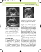
A 8.03 cm
B
image. The anteroposterior diameter is measured from the outer wall to the outer wall (callipers). B: A sagittal image of the same aneurysm, demonstrating the calliper positions to measure the aneurysm in this plane. Note that in this example the diameter measured in the transverse image (A) is larger, due to obliquity.
the longitudinal plane, the aorta is sometimes seen to curve gently in a slight convex direction as it lies on the lumbar spine.
Abnormal appearance
The aorta appears abnormally enlarged, as seen on the B-mode image in Figure 11.6. The shape of the aneurysm can vary (Fig. 11.4). Marked kinking of the posterior wall at the level of the proximal neck can occur due to elongation of the aorta, which can be mistaken as an atherosclerotic stenosis. If the aneurysm deflects in an anterior direction, it can be very difficult to demonstrate the level of the renal arteries, and the proximal segment of the
Figure 11.7 A transverse image of an aortic aneurysm demonstrates a localized area of thrombus liquefaction (L), which may be confused with a dissection. Large areas of thrombus (arrows) separate the area of liquefaction from the flow lumen (FL).
abdominal aorta may become tortuous, which is sometimes described as a ‘swan neck’ appearance.
Thrombus may be imaged as concentric layers with differing degrees of echogenicity depending on the age and organization. Sometimes localized lique- faction of the thrombus can occur, which appears as hypoechoic areas within the thrombus. This appear- ance can be confused with a dissection, although there is usually a thick layer of thrombus separating the liquefied region and the flow lumen (Fig. 11.7). A dissection may be undetected, as blood that has leaked into the wall may be mistaken for mural thrombus. In a full dissection, flow will be observed in the false flow lumen, which is separated from the true lumen by a flap of intima and, sometimes, media (Fig. 11.8).
Inflammatory aneurysms demonstrate a hypo- echoic area of ill-defined fibrosis around the aorta on the B-mode image, but this appearance can be con- fused with the presence of periaortic lymph nodes. The ultrasound diagnosis of a leaking aneurysm is extremely difficult, although it is sometimes possi- ble to identify areas of fresh blood or hematoma as hypoechoic areas associated with the aneurysm in the retroperitoneal space. This type of assessment should be carried out by an experienced sonogra- pher; however, other imaging techniques, such as CT and MRI, are better suited for this investigation.
7.47 cm 7.52 cm
A large abdominal aortic aneurysm is shown from two different imaging planes. A: Transverse
Figure 11.6
