
Page 158 - Libro vascular I
P. 158
Chap-11.qxd 29~8~04 14:49 Page 149
aneurysms. Postoperative recovery is usually very quick, with some patients going home within 3 to 5 days. However, not all aneurysms are suitable for endovascular repair. This can be due to aneurysm tortuosity, excessive proximal neck diameter, limited proximal neck length, severe iliac artery disease and marked iliac artery tortuosity. Although endovas- cular repair appears much less traumatic for the patient, a number of complications are possible, including endoleak. An endoleak occurs when blood leaks into the aneurysm sac from the graft or from another source, such as a lumbar or inferior mesen- teric artery. In this situation the aneurysm sac can continue to expand and rupture (van Marrewijk et al 2002). The different types of endoleak and their ultrasound appearances are discussed later in this chapter. Some devices have been withdrawn from use due to problems such as hook fractures and structural failure. At the time of writing, the long- term durability and outcome of endovascular repair compared to conventional open repair is unknown and is the subject of ongoing trials in Europe and the United States.
ANEURYSM SHAPES AND TYPES
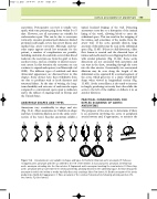
Aneurysms vary considerably in shape and size (Fig. 11.4). Most aneurysms are fusiform in shape and there is uniform dilation across the entire cross- section of the vessel. Saccular aneurysms exhibit a
ABCDEFGHI
TL FL TL
Figure 11.4 Aneurysms are very variable in shapes and types. A: Fusiform infrarenal aortic aneurysm. B: Tortuous elongated aortic aneurysm with the sac shifted to the left of the midline. C: Saccular aortic aneurysm. D: Infrarenal aortic aneurysm extending into the iliac arteries. E. Suprarenal aortic aneurysm involving the renal arteries. F: Dissecting aortic aneurysm with a tear between the intima and media allowing blood into the subintimal space. G: Dissecting aortic aneurysm in which the intima or media has fully dissected, creating a false flow lumen. H: Double aneurysm of the aorta producing a ‘dumb-bell appearance’. I: False aneurysm of the common femoral artery following arterial puncture. (TL, true lumen; FL, false lumen.)
DUPLEX ASSESSMENT OF ANEURYSMS
149
typical localized bulging of the wall. Dissecting aneurysms occur due to a disruption of the intimal lining of the vessel, allowing blood to enter the subintimal space. This can result in the stripping of the intima, and sometimes of the media, from the artery wall. If the aorta partially dissects, large amounts of thrombus may be seen in the subintimal space (Fig. 11.4F). If there is a full dissection, a false flow lumen is created and the dissected layer of intima and media may be seen flapping freely in time with arterial pulsation (Fig. 11.4G). Some aortic dissections are not associated with aneurysms and can start in the chest, extending through the aorta into the iliac arteries. Occasionally, two aneurysmal dilations may be seen along the length of the abdominal aorta, separated by a normal segment of the aorta, which gives rise to a classic ‘dumb-bell’ shape when viewed in longitudinal section (Fig. 11.4H). As the aorta dilates, it also tends to increase in length, producing tortuosity that often shifts the aorta to the left of the midline or deflects it in an anterior direction.
PRACTICAL CONSIDERATIONS FOR DUPLEX SCANNING OF AORTIC ANEURYSMS
The purposes of the scan are to determine if there is an aneurysm involving the aorta or peripheral arterial system and, if appropriate, to monitor the
