
Page 162 - Libro vascular I
P. 162
Chap-11.qxd 29~8~04 14:50 Page 153
DUPLEX ASSESSMENT OF ANEURYSMS
153
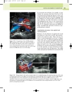
be measured in the AP plane. It is possible to assess whether the aneurysm starts below the level of the renal arteries by measuring the diameter of the aorta at this level with the aid of color flow imaging. With the transducer in a transverse plane, positioned at the upper abdomen, the right renal artery is normally seen dividing from the aorta at a 10 o’clock position and the left renal artery from a 4 o’clock position (Fig. 11.10). In practice, they can be very difficult to image, especially if the aneurysm is tortuous or pro- jecting upward or kinked.
Longitudinal scan plane, from sagittal and coronal positions
Estimates of aortic diameter in sagittal and coronal scan planes are often more accurate than measure- ments in the transverse plane. This is because it is easier to avoid measurement errors due to oblique views of the aorta. To find the maximum diameter of the aorta, the transducer should be swept laterally across the aorta until the widest point can be seen (Figs 11.6 and 11.11). The coronal imaging plane should also be used for measuring the lateral diame- ter of the aneurysm, as some aneurysms are larger in the lateral than in the AP dimension. The length of the aneurysm sac should also be measured in the
S
A LR
V
RR
Figure 11.10 The positions of the left (LR) and right (RR) renal arteries are shown in this transverse image of the aorta (A). The diameter of the aorta is normal at the level of the renal arteries. The left renal vein (V) can be seen crossing over the aorta at the same level as the renal arteries. The SMA (S) is also seen in this image.
SMA
C
D
RV
E
B
A
Figure 11.11
Measurements of an aortic aneurysm made in the longitudinal plane. The maximum diameter of the aorta is measured across line A. The length of the aneurysm sac is measured along line B. The distance from the SMA origin to the approximate level of the renal arteries is measured along line C. The left renal vein (RV) can also be seen in transverse section as it crosses over the aorta at the level of the renal arteries. The distance from the SMA origin to the aneurysm sac is measured along line D. The diameter of the aorta between the aneurysm and the level of the renal arteries is measured across line E.
