
Page 163 - Libro vascular I
P. 163
Chap-11.qxd 29~8~04 14:50 Page 154
154
PERIPHERAL VASCULAR ULTRASOUND
longitudinal scan plane (Fig. 11.11). The presence and thickness of thrombus can sometimes be difficult to assess on a poor B-mode image. Color flow imaging can be useful for demonstrating the lumen, which may be small in the presence of a large thrombus load.
Distance between the renal arteries and
the upper limit of the aneurysm
When requested, the distance between the renal arteries and upper limit of the aneurysm can be measured. In practice, this can be an extremely diffi- cult or virtually impossible assessment to make. First, the presence of the aneurysm may obscure views of the upper abdominal aorta. Second, the renal arter- ies cannot usually be imaged with the probe in the longitudinal direction required to make this meas- urement. However, the position of the renal arteries can be estimated by identifying the SMA in the longitudinal plane, as the renal arteries should lie approximately 1.5cm below the SMA origin (Fig. 11.11). Accessory renal arteries may arise well below this point. The left renal vein can act as another use- ful landmark, if it is found to be at the level of the renal arteries in a transverse image (Fig. 11.10). Turning the transducer into a longitudinal plane, it is possible to identify the renal vein as it crosses over the top of the aorta (Fig. 11.11). Other imaging techniques, such as CT, MRI or arteriography, are generally used to identify the position of the renal arteries in large aneurysms, especially with the increasing use of endovascular devices to repair aneurysms.
LIMITATIONS AND PITFALLS OF AORTIC
ANEURYSM SCANNING
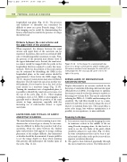
The main limitation of aortic scanning is poor visu- alization due to bowel gas or obesity. It can some- times be difficult to define the posterior wall of an aneurysm, when the tissue between the lumbar spine and posterior wall appears to merge, making placement of the calliper difficult. Any limitations or doubts should be documented. A major pitfall is to set the image depth too deep when scanning thin patients and misinterpret the lumbar spine as the aorta (Fig. 11.12).
V
A
S
In this image, the scanning depth has been set too deep in a thin patient, and the lumbar spine
Figure 11.12
(S) has been mistaken for an aneurysm. The aorta (A) is of normal diameter. The vena cava (V) can be seen to the right of the aorta.
SURVEILLANCE OF ENDOVASCULAR ANEURYSM REPAIR
Duplex ultrasound is an accurate method for the detection of endoleaks following endovascular repair (McLafferty et al 2002). It is important to optimize the scanner controls so that the system is sensitive to detecting low-velocity flow. This can be achieved by reducing the PRF to 1–1.5 kHz and increasing color sensitivity. The wall filter should be set to a mini- mum level and write zoom used to image the area of interest to maintain frame rate. This can produce a noisy color image, but without optimization it is possible, in our experience, to miss endoleaks.
Scanning technique
1. Itiseasiesttostartthescanbyimagingtheaorta in transverse section in the middle of the sac using B-mode imaging alone. At this level it is usual to see the two limbs of the graft, which usually lie adjacent to each other (Fig. 11.13A). In some circumstances they can be seen to spi- ral around each other as the probe is moved in a superior or inferior direction.
2. The graft is then followed proximally through the sac in transverse section. The bifurcation of
