
Page 164 - Libro vascular I
P. 164
Chap-11.qxd 29~8~04 14:50 Page 155
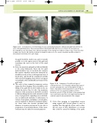
AB
Figure 11.13 A: A transverse color flow image of a successfully deployed aortic endovascular graft taken below the level of the graft bifurcation. Flow is demonstrated in both graft limbs, but there is no flow in the aneurysm sac.
B: Longitudinal color flow image of an endovascular graft. In this example the limbs are lying on top of each other, but in many cases they lie side by side, as shown in example A, where use of a coronal scan plane would be needed to demonstrate both limbs in the same image.
the graft should be clearly seen, and it is usually possible to see the upper extent of the graft and sometimes the aorta at the level of the renal arteries.
3. Next, the aneurysm and graft are followed distally to the aortic bifurcation, where the two graft limbs should be seen to run down the common iliac arteries. Anechoic areas in the aneurysm sac should be noted, as these could represent areas of blood flow, and should be scrutinized carefully with color flow imaging. Some devices can appear very pulsatile, with considerable movement of the graft walls.
4. The aorta is then scanned in transverse section using color flow imaging. There should be color filling of the graft but no flow visible in the sac outside the device (Fig. 11.13A). The maximum diameter of the aneurysm sac should be recorded so that any changes in size can be assessed on serial scans. A progressively expanding scan could indi- cate an undetected endoleak or endotension. A variety of scan planes, including a coronal plane, may be required to obtain the maximum diame- ter. Some units also measure proximal neck diameter to monitor any increase in size due to progression of aneurysmal disease. Other meas- urements may be taken for research purposes.
III
II
II
Figure 11.14
DUPLEX ASSESSMENT OF ANEURYSMS
I III
155
I
A diagram of the different types of endoleak that can occur following endovascular repair of
an aortic aneurysm (see text for description). A: Type I, failure of proximal or distal anastomotic seals. B: Type II, perfusion of the sac via patent lumbar or inferior mesenteric arteries. C: Type III, failure of the modular limb seal or perforation and tears in the graft material. Type IV endoleak cannot be demonstrated in this example.
5. Color flow imaging in longitudinal section using sagittal and coronal planes is used to examine flow through the device and to iden- tify any areas of flow disturbance or stenosis that could be caused by kinking of the graft,
