
Page 166 - Libro vascular I
P. 166
Chap-11.qxd 29~8~04 14:50 Page 157
Type II Collateral endoleaks involve some filling of the sac via lumbar vessels or the inferior mesenteric artery or accessory renal arteries (Fig. 11.15B and C). They can be difficult to detect with ultrasound. Many type II endoleaks will seal spontaneously after a month or two (Veith et al 2002). Persistent leaks may require embolization.
Type III Leaks between the modular limb and main body of the graft or tears in the graft (Fig. 11.15D). These types of leaks are less commonly seen.
Type IV Thought to occur due to graft porosity or ‘sweating’ of graft material, leading to progressive increase in sac size within the first month. It is not possible to image this type of leak in real-time, but serial surveillance scans may show a progressive increase in the diameter of the aneurysm sac.
In addition, the concept of endotension has been defined. This is described as a persistent or recur- rent pressurization of the sac without a visualized endoleak. This can lead to expansion of the sac and potential rupture. The causes of endotension are uncertain, but it has been suggested that the sac is pressurized by mechanisms such as excessive pulsa- tion of the graft, osmosis into the sac or transmis- sion of pressure through the thrombus. In practice, some cases of endotension could be due to a very small, undetected endoleak.
The management of endoleaks remains unclear, but there is evidence to suggest that type I and III endoleaks should be treated, as they are more likely to be associated with an increase in sac size and potential rupture.
Assessment of aneurysms excluded
by covered stents
Flow can be excluded in aneurysms involving other areas of the arterial circulation by inserting a cov- ered stent across the region of the aneurysm. The proximal and distal ends of the stent are positioned above and below the aneurysm. These types of stents are most commonly used in the iliac arteries to exclude true aneurysms. They can also be used to exclude flow into false aneurysms following vessel perforation or rupture during angioplasty. They are occasionally used in the subclavian arteries.
I
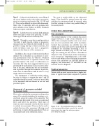
This longitudinal image of a distal aortic aneurysm (A) and right common iliac artery (I)
demonstrates two large iliac artery aneurysms (seen between arrows).
DUPLEX ASSESSMENT OF ANEURYSMS
157
The stent is usually visible on the ultrasound image. Flow should be assessed across the stent and then the aneurysm should be scanned, using low-flow settings, to detect any possible filling of the aneurysm sac.
OTHER TRUE ANEURYSMS
Iliac aneurysms
The normal diameter of the common iliac artery ranges between 1.1 and 1.4cm (Johnston et al 1991). Iliac aneurysms usually occur as an exten- sion of, or in association with, aortic aneurysms. Isolated iliac aneurysms are relatively rare, but rupture can be fatal, and elective repair should be considered for aneurysms measuring 3.5 cm or larger (Santilli et al 2000) (Fig. 11.16). The tech- nique for scanning the iliac arteries is described in Chapter 9. The measurement of iliac aneurysms is more accurate in a longitudinal plane, as it is diffi- cult to avoid oblique planes if imaging in transverse section. Iliac aneurysms are clinically difficult to diagnose, and ultrasound, CT and MRI are the methods used for diagnosis.
Popliteal aneurysms
Patients with an aortic aneurysm have a higher inci- dence of popliteal aneurysms compared to patients
A
Figure 11.16
