
Page 168 - Libro vascular I
P. 168
Chap-11.qxd 29~8~04 14:50 Page 159
DUPLEX ASSESSMENT OF ANEURYSMS
159
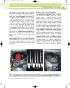
backward and forward through a hole in the arterial wall into the surrounding tissue, forming a flow cav- ity in the tissue adjacent to the artery. The false lumen often contains thrombus, which may be lay- ered. False aneurysms can increase in size over time. Color flow imaging should be used to confirm flow in the false lumen. The color flow image typically demonstrates a high-velocity jet originating from the defect in the artery wall, which is associated with a swirling pattern inside the false lumen, similar to the ‘yin-yang’ sign. Spectral Doppler usually demonstrates an equal forward flow and reverse flow component to the arterial jet as flow enters the false aneurysm during systole and exits during dias- tole (Fig. 11.18). The audible Doppler signal is very characteristic, with high-frequency Doppler shifts heard in the forward and reverse phases across the neck.
The common femoral artery is the main vessel in which false aneurysms occur, as it is the commonest site for catheter access. False femoral aneurysms may be very large, and bleeding into the retroperitoneal cavity can be a serious complication, leading to shock and death.
FL
A
AB
T
A
Scanning false femoral aneurysms
The patient should lie as flat as possible. The proce- dure should be started by scanning the common femoral artery in transverse section. A mid- frequency 5MHz, or broad-band equivalent, flat linear array transducer will usually provide an ade- quate image. However, in some cases an abdominal curved array transducer may be required, especially if the patient is obese or if the puncture has been very high. In addition, areas of hematoma lying over the vessel, associated with the puncture site, can make the imaging difficult. The common femoral artery should be identified and scanned along its length in transverse section using color flow imag- ing. The proximal few centimeters of the superficial femoral artery and profunda femoris artery should also be examined, as low punctures can result in false aneurysms of these vessels. A potentially confusing situation can occur if the inferior epigastric artery, a superficial branch of the common femoral artery, runs close to an area of hematoma or swelling, as this might be mistaken for a small leak. Spectral Doppler recordings taken from the superficial epigastric
Figure 11.18 A: A transverse image of a false femoral artery aneurysm and corresponding spectral Doppler waveform from the communicating jet. The arrow indicates the arterial jet between the femoral artery (A) and the false lumen (FL). There is marked perivascular tissue vibration associated with the arterial jet in this example. B: The false aneurysm was successfully thrombosed (T) by ultrasound guided compression.
