
Page 167 - Libro vascular I
P. 167
Chap-11.qxd 29~8~04 14:50 Page 158
158
PERIPHERAL VASCULAR ULTRASOUND
used to demonstrate the size of the flow lumen if the B-mode imaging is poor. Occluded popliteal aneurysms are demonstrated by an absence of color flow in the lumen. The popliteal vein should also be assessed for patency. Baker’s cysts have a typical appearance of a tail trailing from the main body of the cyst to the joint capsule, and they have a hypo- echoic appearance due to the synovial fluid inside (see Ch. 13).
Femoral artery aneurysms
True femoral artery aneurysms occur less frequently and are usually associated with aneurysmal disease elsewhere. However, aneurysmal dilations can occur where graft anastomoses have been performed.
FALSE ANEURYSMS
False aneurysms, also known as pseudo-aneurysms, primarily occur following arterial puncture for catheter access, due to poor control of arterial bleed- ing following the procedure. This is usually due to insufficient pressure being applied over the puncture site or pressure being applied for too short a time. They may also occur following trauma. Blood flows
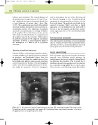
V
PA
without such aneurysms. The normal diameter of the popliteal artery ranges between 0.4 and 0.9 cm, and a popliteal aneurysm is frequently classified as a vessel diameter of greater than 1.5 cm (Fig. 11.17). The complications associated with popliteal aneurysms include embolization, acute occlusion or, occasionally, rupture. The symptoms of popliteal aneurysms can include pain or a feeling of fullness in the popliteal fossa. Sometimes patients present with a deep vein thrombosis due to compression of the popliteal vein by the aneurysm. Ultrasound is the primary technique for the diagnosis of popliteal aneurysms. However, a major diagnostic pitfall is the misdiagnosis of a Baker’s cyst as a popliteal aneurysm.
Scanning of popliteal aneurysms
Using a 5 MHz, or broad-band equivalent, flat lin- ear array transducer, the popliteal artery is examined in transverse and longitudinal planes from the popliteal fossa and from the medial aspect of the lower thigh in the adductor canal, as some aneurysms can be located above the knee. B-mode imaging is used to assess the size, length and amount of throm- bus within the aneurysm. Color flow imaging is
V
PA
T
AB
Figure 11.17 A: A transverse image of a popliteal artery aneurysm (PA) containing thrombus (T) below the arrow. The popliteal vein (V) is compressed in this example. B: A longitudinal image demonstrating thrombus in the lumen. The popliteal vein is seen superficial to the artery.
