
Page 232 - Libro vascular I
P. 232
Chap-14.qxd 29~8~04 14:56 Page 223
GRAFT SURVEILLANCE AND PREOPERATIVE VEIN MAPPING FOR BYPASS SURGERY
223
removed and the dots joined up with a continuous line using a permanent felt-tipped marker pen.
The second technique involves assessing the vein in transverse section for its size and position, and then marking the vein with the transducer turned into a longitudinal plane and the vein imaged in this direction. A dot is placed against the end of the transducer to correspond to the position and direc- tion of the vein. This technique is more difficult in terms of imaging, as it is easy to ‘slip off’ the image of the vein and follow a tissue plane, but it does seem to produce a more accurate map of the vein. Once the position of the vein has been marked using this technique, it is wise to run down the length of marking with the transducer in transverse section to ensure that the dots follow the main trunk, in case a smaller side branch in the lower leg has been accidentally followed using this method.
Problems encountered during vein mapping
One major practical problem of vein mapping is trying to mark the position of the vein with a felt- tipped pen through the ultrasound gel, as the pen quickly becomes clogged with gel and no longer works. Many vascular units have their own personal preferences for overcoming this problem. A char- coal pencil can be useful for dotting in the position of the vein and the dots joined up later with a felt- tipped pen once the gel has been removed. Alter- natively, using small amounts of gel and frequently wiping the gel away from the probe edge before marking prevent the pen tip from becoming satu- rated in ultrasound gel. It is also useful to place a non-sterile probe cover over the transducer when vein mapping, as this prevents the probe from becoming covered in ink from the marker pen.
If the patient has very poor muscle volume and the superficial tissues are baggy, it can be very dif- ficult to ensure that the marking is accurate. If in any doubt, tell the surgeon. In obese patients, the vein may be very deep, and it can be difficult to mark it in the correct incision plane.
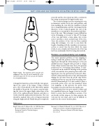
A
X X
V
X
X V
Figure 14.24
B
Two methods can be used for vein mapping. A: The vein (V) can be marked (X) in the
transverse plane. B: The vein can also be marked in a longitudinal plane.
is imaged in transverse section with the vein appear- ing in the center of the image. Using a marker pen, a dot is then placed on the skin surface against the middle of the probe. It is easier to start by mark- ing the vein in the upper thigh, rather than at the level of the saphenofemoral junction and then to work toward the saphenofemoral junction. The vein should be marked with a dot at frequent intervals along the thigh and calf. Finally the gel is completely
References
Bagi P, Schroeder T, Sillesen H, et al 1989 Real time B-mode mapping of the greater saphenous vein. European Journal of Vascular Surgery 3(2):103–105
Brennan J A, Walsh A K, Beard J D, et al 1991 The role of simple non-invasive testing in infrainguinal graft surveillance. European Journal of Vascular Surgery 5(1):13–17
