
Page 81 - Libro vascular I
P. 81
Chap-06.qxd 29~8~04 14:41 Page 72
72
PERIPHERAL VASCULAR ULTRASOUND
A
B
S
S
D
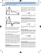
Figure 6.9 Pulsatility index (A & B) and resistance index
M
DE
M
quantify the degree of pulse wave damping at dif- ferent measurement sites. It is defined as the max- imum height of the waveform, S, minus the minimum diastolic, D (which may be negative), divided by the mean height, M, as shown in Figure 6.9A and B:
(6.4)
Damped flow beyond significant disease will have a lower PI value than a normal pulsatile waveform.
Pourcelot’s resistance index
PI S D M
The resistance index (RI) was first used on com- mon carotid waveforms as an indicator of the peripheral resistance and has also been used to study neonatal cerebral hemodynamics. It is defined as follows (Fig. 6.8A):
(6.5)
where E is end diastolic velocity. The value of RI can be calculated by the scanner and displayed on the screen.
Spectral broadening
RI S E S
There have been several definitions of spectral broadening (SB) described over the years in an attempt to quantify the spread of frequencies pres- ent within a spectrum. One such definition is as follows:
SB fmax fmin f max
(6.6)
Increased SB indicates the presence of arterial disease but can, to some extent, also be introduced by the scanner itself, as in ISB (described above).
Pulse wave velocity
The pressure pulse and the associated velocity wave travel along the vessel at a different speed from the flowing blood. The speed at which the pulse is transmitted along the vessel depends on the elas- ticity of the vessel wall. For example, the pulse will travel much faster down the stiff-walled artery of a
(A) can be calculated from the peak systolic, S, minimum diastolic, D, end diastolic, E, and the mean velocity (or frequency), M, shown here on two different waveforms.
as the Doppler system is unable to differentiate between the noise and the Doppler signals.
WAVEFORM ANALYSIS
As well as the blood velocity and flow changing with the presence of significant disease, the shape of the waveform will also be altered (as discussed in Ch. 5). The waveform may indicate whether the disease is proximal or distal to the site at which the Doppler signal is obtained. Over the years, several researchers have attempted to quantify these changes in wave- form shape by defining various indices, and many modern scanners incorporate facilities to calculate such quantities, some of which are listed below.
Pulsatility index
The pulsatility index (PI) is probably the most commonly used of all the indices. It can be used to
