
Page 79 - Libro vascular I
P. 79
Chap-06.qxd 29~8~04 14:41 Page 70
70
PERIPHERAL VASCULAR ULTRASOUND
velocity measurements:
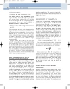
● Always set the angle of insonation to 60°
This ensures that any error in alignment of the angle correction cursor only leads to a moderate error in the velocity estimate (Fig. 6.7) and that the errors caused by ISB are kept similar between measurements. However, it can be difficult to insonate all vessels at a fixed 60° angle.
● Always select as small an angle of insonation as possible
This ensures that any error in the alignment of the angle correction cursor produces as small an error in velocity estimation as possible. The error due to ISB will also be minimized. However, this error will be different for measurements made at differ- ent angles of insonation. This makes comparisons between measurements made at different angles less meaningful.
Doppler criteria developed over the years may not have been produced with a full understanding of all these possible sources of error. Different mod- els of ultrasound system may produce different results for the same blood flow. However, despite these sources of error, velocity measurements have been successfully used to quantify vascular disease for the past two decades. A greater understanding of the sources of error in velocity measurement may lead to improvements in accuracy.
Other potential sources of error in
maximum velocity measurements
Figure 3.7 has already shown how high-pass filters can be used to remove unwanted signals. The high-pass filter setting will not affect the peak sys- tolic velocity measurements, but the shape of the peak velocity envelope (the outline of the spec- trum) may be affected if the filter is set so high that it removes the diastolic flow. This would lead to an incorrect finding that the end diastolic velocity is zero. Figure 3.14 shows how aliasing will lead to an underestimation in the mean velocity and the maximum velocity due to the incorrect estimation of the high frequencies present within the signal. Noise may be introduced into the Doppler signal, especially if the signal is recorded at depth, requiring
significant amplification. The maximum Doppler fre- quency may be difficult to define in the presence of high levels of noise.
MEASUREMENT OF VOLUME FLOW
Volume flow is a potentially useful physiological parameter (see Fig. 5.4) that can be measured using ultrasound, although it involves several pos- sible sources of error (Evans & McDicken 2000). An estimation of the volume flow of blood can be made if the cross-sectional area of the vessel and the velocity of the blood through the vessel are known. Ultrasound scanners usually have the facility to perform volume flow measurements by enabling the sonographer to measure diameter or cross-sectional area from the image and then to measure the TAV from the Doppler spectrum, cal- culating the flow as follows:
Flow cross-sectional area TAV (6.2)
The most straightforward method of obtaining the vessel cross-sectional area is to measure the ves- sel diameter (d) and calculate the area as follows:
A A p p d d 2 2 4
(6.3)
Some scanners also allow the sonographer to outline the circumference of the vessel, imaged in transverse section, using a cursor. This method tends to be less reliable as it requires a steady hand and a good image of the lateral walls of the vessel. The cross-sectional area can then be multiplied by the TAV to give the flow, as shown in Figure 6.6B.
Sources of error in vessel diameter
measurement
Errors in either the velocity measurement or the diameter measurement will introduce errors into the estimation of volume flow. As flow is propor- tional to the cross-sectional area of the vessel, which in turn depends on the square of the radius, any error in the diameter will produce a fractional error in the flow measurement that is double the fractional error in the radius. The possible sources
