
Page 95 - Libro vascular I
P. 95
Chap-08.qxd 1~9~04 16:41 Page 86
86
PERIPHERAL VASCULAR ULTRASOUND
ANATOMY
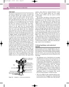
The brain is supplied by four vessels—the right and left internal carotid and vertebral arteries—and receives 15% of the cardiac output. The term extra- cranial cerebral arteries refers to all the arteries that carry blood from the heart up to the base of the skull. The left and right sides of the extracranial circulation are not symmetrical (Fig. 8.1). On the left side, the common carotid (CCA) and subcla- vian arteries arise directly from the aortic arch, whereas on the right side the brachiocephalic artery, also known as the innominate artery, arises from the aorta and divides into the subclavian artery and CCA. The CCA, which has no branches, divides into the internal and external carotid arteries, but the level of the carotid bifurcation in the neck is highly variable. In approximately 90% of cases, the internal carotid artery (ICA) lies posterolateral or lateral to the external carotid artery (ECA) and, unlike the ECA, has no branches below the skull. The proximal branches of the ECA are the superior thyroid, lingual, facial and maxillary arteries. The carotid artery widens, at the level of the bifurcation, to form the carotid bulb. In some cases, the carotid bulb may only involve the proximal ICA, and not the distal CCA, and the degree of widening of the carotid bulb is quite variable. Within the skull, the distal segment of the ICA follows a curved path, known as the carotid siphon. The most important branch of the ICA is the ophthalmic artery, which supplies the eye. The terminal branches of the oph- thalmic artery, the supratrochlear and supraorbital
arteries, unite with the terminal branches of the ECA. The ICA finally divides into the middle cerebral artery (MCA) and the anterior cerebral artery (ACA).
The posterior circulation of the brain is mainly supplied by the left and right vertebral arteries, via the basilar artery. The vertebral artery is the first branch of the subclavian artery, arising from the highest point of the subclavian arch. At the sixth cervical vertebra, the vertebral artery runs posteriorly to travel upward through the transverse foramen of the cervical vertebrae. It is common for one vertebral artery to be larger than the other, with the left often being larger than the right. The two vertebral arter- ies join, at the base of the skull, to form the basilar artery, which then divides to form the posterior cere- bral arteries. Figure 8.2A shows how the circle of Willis, situated at the base of the brain, joins the cerebral branches of the ICAs and basilar artery via the anterior and posterior communicating arteries. Blood flow to the brain is regulated by changes in cerebrovascular resistance, with carbon dioxide play- ing a major role in vasodilation.
Collateral pathways and anatomical
variants
In the presence of severe vascular disease, the cere- bral circulation has many possible collateral (alterna- tive) pathways, both extracranially and intracranially. Not all of these can be assessed using ultrasound; however, two pathways that can be assessed are the following:
● The ophthalmic artery. The ECAs do not normally supply blood to the brain, but in the presence of severe ICA disease, branches of the ECA can act as important collateral pathways. One important collateral pathway is via the terminal branches of the ECA, communicating with the terminal branches of the ophthalmic artery. This colla- teral pathway can be observed using continuous wave (CW) Doppler to detect reversal of flow in the supraorbital artery, a terminal branch of the ophthalmic artery, as retrograde flow travels from the ECA branches toward the brain.
● The circle of Willis. In the normal circulation, there is little blood flow through the communicating arteries in the circle of Willis, but in the presence
Right
Carotid bulb
Subclavian artery
Brachiocephalic artery (also known as innominate)
Aorta
Anterior cerebral artery Middle cerebral artery
Ophthalmic artery Posterior cerebral artery Basilar artery External carotid artery
Internal carotid artery
Common carotid artery Vertebral artery
Subclavian artery
Figure 8.1 Diagram of cerebrovascular anatomy.
