
Page 96 - Libro vascular I
P. 96
Chap-08.qxd 1~9~04 16:41 Page 87
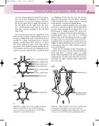
of severe vascular disease they perform an impor- tant role in flow distribution. For example, in the presence of a left ICA occlusion, it is possi- ble for the right ICA to supply blood flow to the left MCA via the right ACA, the ante- rior communicating artery and the left ACA, with flow reversal occurring in the left ACA (Fig. 8.2B).
The vertebral arteries may also supply flow to the MCA via the posterior communicating arteries of the circle of Willis. If the circle is well developed, it is possible for a single extracranial artery to provide adequate cerebral blood flow. However, in about 75% of the population, parts of the circle may be hypoplastic (very small) or absent, making the cir- cle incomplete and therefore preventing the devel- opment of good collateral flow (von Reutern &
ULTRASOUND ASSESSMENT OF THE EXTRACRANIAL CEREBRAL CIRCULATION
87
von Büdingen 1993), but this may only become apparent in the presence of severe disease. Adequate collateral pathways have a better chance to develop in the presence of slowly developing disease.
An unusual collateral pathway can occur when the CCA is occluded and flow in the proximal ECA reverses, being supplied by retrograde flow in an ECA branch, to supply a patent ICA. Severe nar- rowing or occlusion of the proximal subclavian or brachiocephalic artery can result in a collateral pathway that ‘steals’ blood from the brain to supply the arm. In this case, blood will be seen to flow ret- rogradely down the ipsilateral vertebral artery to supply the distal subclavian artery beyond the dis- eased segment (Fig. 8.3). This is known as subcla- vian steal syndrome.
There are few variations in the extracranial cir- culation. In rare cases, the left CCA and subclavian artery may share a common origin or a single trunk. Other anomalies are the left vertebral artery arising directly from the aortic arch and, even more unusually, the right vertebral origin arising from the aortic arch.
Right
A
Right
B
cross-over flow from the right ICA to the left MCA in the presence of a left ICA occlusion.
Anterior communicating artery Anterior cerebral artery
Middle cerebral artery
Internal carotid artery
Posterior communicating artery
Posterior cerebral artery
Basilar artery
Occluded left ICA
Diagram of the circle of Willis. A: Arrows indicate normal flow direction. B: Arrows indicate
Right
Vertebral arteries
Figure 8.2
Occluded proximal subclavian artery
Arrows indicate the direction of collateral flow in subclavian steal syndrome, via reverse flow in the vertebral artery to supply the arm, in the presence of a severe stenosis or occlusion of the proximal subclavian artery.
Figure 8.3
