
Page 98 - Libro vascular I
P. 98
Chap-08.qxd 1~9~04 16:41 Page 89
also demonstrate the presence and direction of flow in the vertebral arteries. No specific preparation is required, but the patient must be capable of lying or sitting still during the examination. The optimal position for scanning the carotid arteries is with the sonographer sitting behind the patient’s head. This allows easy access to the neck and enables the opera- tor to rest the arm on the examination table while performing the scan (Fig. 8.4). Alternatively the sonographer can sit by the side of the patient while resting the arm on the patient’s upper chest. The patient should lie supine on the couch with the head resting on a pillow. The neck should be extended and the head turned in the opposite direction to the side being examined. If the patient has difficulty in breathing or has back problems it may be necessary to sit the patient in a more upright position. If the patient is in a wheelchair (e.g., following a disabling stroke), it may be easier to do the scan in the wheel- chair with the head resting on a pillow for support, preventing unnecessary movement of the patient.
The examination can be performed with a medium- to high-frequency (e.g., broad-band 4–7 or 5–12MHz) flat linear array transducer. The higher the frequency, the better the resolution of the vessel wall structure; however, in some cases the carotid bifurcation lies deep in the neck, requiring a lower frequency transducer for visualization. Blood flow velocities detected in the majority of normal and diseased carotid arteries are reasonably high, so the scanner should be configured to visualize
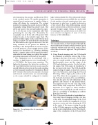
Figure 8.4 The optimal position for scanning the carotid arteries.
ULTRASOUND ASSESSMENT OF THE EXTRACRANIAL CEREBRAL CIRCULATION
89
high-velocity pulsatile flow. Most ultrasound systems have examination presets available that are suitable for the majority of carotid examinations, but it may be necessary to alter these to enable the detection of low-velocity flow when differentiating carotid artery occlusion from a subtotal occlusion. A small spectral Doppler sample volume is usually used to interrogate the carotid arteries, as it allows for more selective investigation of areas of velocity increase or flow disturbance.
Technique
The carotid arteries are best visualized through the sternocleidomastoid muscle, which provides a good ultrasonic window, and this is done using a lateral rather than an anterior approach. The procedure is as follows:
1. Using B-mode imaging only, the CCA should be visualized in transverse section (Fig. 8.5A), starting at the base of the neck. On the right side, it is usually possible to visualize the distal brachiocephalic artery and the origin of the CCA and subclavian arteries. On the left side, the origin of the CCA cannot be visualized as it lies too deep in the chest. The CCA should be scanned along its length, in transverse section, up to the bifurcation, and along the ICA and ECA (Fig. 8.5B) as high up the neck as can be seen. This allows the sonographer to ascertain the level and orientation of the carotid bifurca- tion and also gives the first indications of the presence and location of any arterial disease. The jugular vein lies over the CCA (Fig. 8.5A) and is usually easily compressed. However, it is important not to apply too much transducer pressure when scanning the carotid arteries as there is a possibility of dislodging an embolus from the vessel wall.
2. The CCA is now visualized in longitudinal sec- tion using B-mode imaging, starting at the base of the neck. A longitudinal image of the CCA can be easily obtained by imaging the CCA in transverse section and then, keeping the CCA in the center of the image, rotating the probe so the CCA first appears as an ellipse and finally can be seen in longitudinal section. Prior knowl- edge of the orientation of the ICA and ECA
