
Page 175 - parasitology for medical and clinical laboratoryprofessionals
P. 175
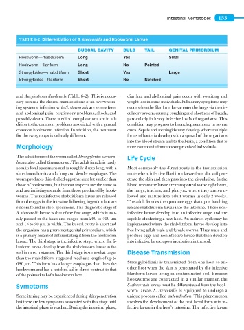
Intestinal Nematodes   155


                    TABLE 6-2  Differentiation of S. stercoralis and Hookworm Larvae

                                              BUCCAL CAVITY       BULB      TAIL       GENITAL PRIMORDIUM
                    Hookworm—rhabditiform     Long                Yes                  Small
                    Hookworm—filariform       Long                No        Pointed
                    Strongyloides—rhabditiform  Short             Yes                  Large
                    Strongyloides—filariform  Short               No        Notched


                   and Ancylostoma duodenale (Table 6-2). This is neces-  diarrhea and abdominal pain occur with vomiting and
                   sary because the clinical manifestations of an overwhelm-  weight loss in some individuals. Pulmonary symptoms may
                   ing systemic infection with S. stercoralis are severe fever   occur when the filariform larvae enter the lungs via the cir-
                   and abdominal pain, respiratory problems, shock, and   culatory system, causing coughing and shortness of breath,
                   possibly death. These medical complications are in ad-  particularly in heavy infective loads of organisms. This
                   dition to the common problems associated with a general     condition may progress to bronchopneumonia in severe
                   common hookworm infection. In addition, the treatment   cases. Sepsis and meningitis may develop where multiple
                   for the two groups is radically different.       forms of bacteria develop with a spread of the organisms
                                                                    into the blood stream and to the brain, a condition that is
                   Morphology                                       more common in immunocompromised individuals.

                   The adult forms of the worm called Strongyloides stercora-  Life Cycle
                   lis are also called threadworms. The adult female is rarely
                   seen in fecal specimens and is roughly 2 mm long, with a   Most commonly the direct route is the transmission
                   short buccal cavity and a long and slender esophagus. The   route where infective filariform larvae from the soil pen-
                   worm produces thin-shelled eggs that are a bit smaller than   etrate the skin and then pass into the circulation. In the
                   those of hookworms, but in most respects are the same as   blood stream the larvae are transported to the right heart,
                   and are indistinguishable from those produced by hook-  the lungs, trachea, and pharynx where they are swal-
                   worms. The noninfective rhabditiform larvae are released   lowed and mature into adult worms in only 2 weeks.
                   from the eggs in the intestine following ingestion but are   The adult females then produce eggs that upon hatching
                   seldom found in stool specimens. The diagnostic stage of     release rhabditiform larvae into the intestine. These non-
                   S. stercoralis larvae is that of the first stage, which is usu-  infective larvae develop into an infective stage and are
                   ally passed in the feces and ranges from 200 to 400 μm    capable of infecting a new host. An indirect cycle may be
                   and 15 to 20 μm in width. The buccal cavity is short and   implemented where the rhabditiform larvae develop into
                   the organism has a prominent genital primordium, which   free-living adult male and female worms. They mate and
                   is a primary means of differentiating it from the hookworm   produce eggs and noninfective larvae that then develop
                   larvae. The third stage is the infective stage, where the fi-  into infective larvae upon incubation in the soil.
                   lariform larvae develop from the rhabditiform larvae in the
                   soil in most instances. The third stage is somewhat larger   Disease Transmission
                   than the rhabditiform stage and reaches a length of up to
                   680 μm. This form has a longer esophagus than does the   Strongyloidiasis is transmitted from one host to an-
                   hookworm and has a notched tail in direct contrast to that   other host when the skin is penetrated by the infective
                   of the pointed tail of a hookworm larva.         filariform larvae living in contaminated soil. Because
                                                                    hookworms are contracted in a similar manner, the
                   Symptoms                                         S. stercoralis larvae must be differentiated from the hook-
                                                                    worm larvae. S. stercoralis is equipped to undergo a
                   Some itching may be experienced during skin penetration   unique process called autoinfection. This phenomenon
                   but there are few symptoms associated with this stage until   involves the development of the first larval form into in-
                   the intestinal phase is reached. During the intestinal phase,   fective larvae in the host’s intestine. The infective larvae