
Page 277 - parasitology for medical and clinical laboratoryprofessionals
P. 277
Laboratory Procedures for Identifying Parasitic Organisms and Their Ova 257
in fixed or preserved samples are destroyed in the
process. New preservatives have been developed due to
Occupational Safety and Health Administration (OSHA)
regulations requiring safety in disposal of materials. For-
merly PVA emitted toxic fumes from formalin and also
contained mercury, so these have been largely replaced
by environmentally safe zinc and copper-based PVA
(polyvinyl alcohol). The new fixative kits to preserve
specimens serve to provide for adequate studies of mor-
phology. In addition, they, do not interfere with staining
procedures or in the subsequent performance of other Delmar/Cengage Learning
immunological tests (see Figure 12-1). These kits con-
taining specimens should also be shipped in a leakproof
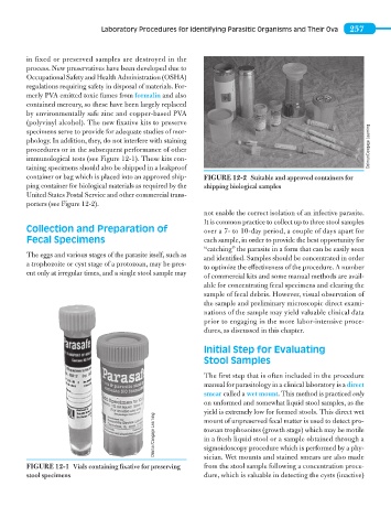
container or bag which is placed into an approved ship- FIGURE 12-2 Suitable and approved containers for
ping container for biological materials as required by the shipping biological samples
United States Postal Service and other commercial trans-
porters (see Figure 12-2).
not enable the correct isolation of an infective parasite.
It is common practice to collect up to three stool samples
Collection and Preparation of over a 7- to 10-day period, a couple of days apart for
Fecal Specimens each sample, in order to provide the best opportunity for
“catching” the parasite in a form that can be easily seen
The eggs and various stages of the parasite itself, such as and identified. Samples should be concentrated in order
a trophozoite or cyst stage of a protozoan, may be pres-
to optimize the effectiveness of the procedure. A number
ent only at irregular times, and a single stool sample may
of commercial kits and some manual methods are avail-
able for concentrating fecal specimens and clearing the
sample of fecal debris. However, visual observation of
the sample and preliminary microscopic direct exami-
nations of the sample may yield valuable clinical data
prior to engaging in the more labor-intensive proce-
dures, as discussed in this chapter.
Initial Step for Evaluating
Stool Samples
The first step that is often included in the procedure
manual for parasitology in a clinical laboratory is a direct
smear called a wet mount. This method is practiced only
on unformed and somewhat liquid stool samples, as the
yield is extremely low for formed stools. This direct wet
Delmar/Cengage Learning tozoan trophozoites (growth stage) which may be motile
mount of unpreserved fecal matter is used to detect pro-
in a fresh liquid stool or a sample obtained through a
sigmoidoscopy procedure which is performed by a phy-
from the stool sample following a concentration proce-
FIGURE 12-1 Vials containing fixative for preserving sician. Wet mounts and stained smears are also made
stool specimens dure, which is valuable in detecting the cysts (inactive)