
Page 304 - parasitology for medical and clinical laboratoryprofessionals
P. 304
284 CHAPTER 12
units. This process is necessary for each objective (10 3, 0.0 0.1 0.2 0.3 0.4
40 3, and oil immersion objectives). To determine how
the ocular units correspond to actual millimeters in size,
a stage micrometer must be used to calibrate the ocular
micrometer. If a different microscope is being used other Delmar/Cengage Learning
than the one that is properly calibrated, a calibration for
0 1 2 3 4 5 6 7
that microscope must be performed, and there are subtle
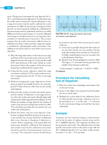
differences between microscopes. A correctly calibrated FIGURE 12-16 Stage micrometer and ocular
microscope is crucial because size is an important char- micro meter superimposed
acteristic for identification of parasites. This section
assumes that an ocular micrometer disk has been in-
be posted on the side of the microscope for quick
stalled in one of the oculars and that a stage micrometer
reference.
is available for calibrating the ocular micrometer. This
a. Look as far as possible along both sides until you
calibration should be done for each of the microscope’s
see two lines exactly over one another. On this
objectives.
scale, the numbers that coincide are 70 and 0.4.
1. Place the stage micrometer on the microscope stage At higher magnifications it may be necessary due
and focus on the micrometer scale, until you can dis- to the thickness of the lines.
tinguish between the large (0.1 mm) and the small b. Divide 0.4 by 70 and multiply the result by 1000.
(0.01 mm) divisions of the scale. Install an ocular The figure, 5.7 (rounded down), provides the
micrometer disk in the eyepiece of the microscope number of microns per ocular unit.
by placing it underneath the eyepiece lens. In this example, 1 ocular unit 5 0.40/70 3 1000 5
2. Using the low power objective, adjust the stage 5.7 microns
micrometer so that the “0” line on the ocular microm-
eter is superimposed with the “0” line on the stage
micrometer. Procedure for Calculating
3. Without changing the stage adjustment, find a Size of Organism
point as distant as possible from the two superim-
1. Place the ocular lens containing a micrometer disc
posed “0” lines where two other lines are also exactly
on the microscope.
superimposed.
2. Focus on the object to be measured and determine
4. Determine the number of ocular micrometer spaces
the size in ocular units.
and the number of millimeters on the stage microm-
eter where the ocular micrometer directly aligns with 3. Multiply the ocular units by the calibration factor
a division line of the stage micrometer (Figure 12-16). for that specific microscope, objective, and ocu-
Divide the number of stage units by the number of lar micrometer (i.e., 1 ocular unit 5 5.7 microns
ocular units and then multiply the results by 1000. for the microscope being used and that has been
This calculation provides the micrometers for one calibrated).
ocular unit on low power.
5. Follow the above steps for each objective. Cali- Example
bration readings should be posted on each micro-
scope and the microscope should be recalibrated A parasite cyst was measured using an ocular microm-
after every cleaning or changing of objectives or eter in the eye piece of a phase contrast scope and its
oculars. Before preparing a wet mount slide, the 40 3 darkfield objective. The organism was three ocu-
microscope should be calibrated. The objectives lar micrometer units wide. The calibration factor for that
and oculars used for the calibration procedure specific micrometer used on the phase scope with the
should be used for all measurements on the mi- 40 3 darkfield objective is 5.7 um. three ocular microm-
croscope. The calibration factors should always eter units 3 5.7 um 5 17.5 um wide.